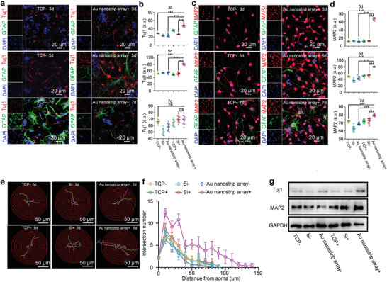
Figure 3.

Au nanostrip array‐based wireless device accelerates neuronal differentiation of NSCs at the protein level. a) Confocal microscopy images of NSCs seeded on TCP or Au nanostrip array and cultured without or with the rotating magnetic field (300 rpm) for 3, 5, or 7 days. Tuj1 and GFAP were stained red and green, respectively. Cell nuclei were stained blue with 4',6‐diamidino‐2‐phenylindole (DAPI). b) Quantitative mean immunofluorescence intensity of Tuj1. c) Confocal microscopy images of NSCs seeded on TCP or Au nanostrip array and cultured without or with the rotating magnetic field (300 rpm) for 3, 5, or 7 days. MAP2 and GFAP were stained red and green, respectively. Cell nuclei were stained blue with DAPI. d) Quantitative mean immunofluorescence intensity of MAP2. b,d) At least 30 cells were analyzed in each group using Image J software. The data are presented as mean ± standard deviation; ns p > 0.05, ***p < 0.001. e) Images of 2D‐reconstructed neuronal morphologies that were traced and visualized based on the Tuj1 protein expression of NSCs seeded on TCP, Si, or Au nanostrip array, and cultured without or with the rotating magnetic field (300 rpm) on day 5. f) Sholl analysis of the neurite complexity of the 2D‐reconstructed neuronal morphologies presented in e). Neurites of ten neurons on each group were randomly measured. g) Western blot analysis of the Tuj1 and MAP2 protein expression of NSCs seeded on TCP, Si, or Au nanostrip array, and cultured without or with the rotating magnetic field (300 rpm) on day 5. Glyceraldehyde 3‐phosphate dehydrogenase (GAPDH) was used as the housekeeping gene.
