Figure 1.

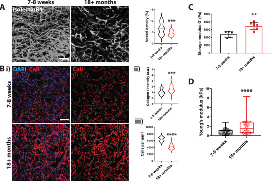
An increase in tissue stiffness correlates with a decrease in vascular density in aged kidneys. A) Representative 3D projection of cleared kidney tissue. Vasculature was visualized using isolectinB4. Scale bar is 50 µm. Quantification of vascular density. Vessel volume was quantified and normalized to the total volume of the imaged area. N = 4 animals with a total of 28 images. Bi) Representative images of Collagen I staining of a tissue section of aged and young mouse kidneys (DAPI in blue and Collagen I in red). Scale bar is 50 µm. ii) Quantification of relative collagen levels normalized to DAPI shows an increase in Collagen I deposition in aged kidneys iii) and a decrease in cell density. N = 3 kidneys with a total of 30 images were analyzed. C) Tissue rheology of young and aged mouse kidneys shows an increase in overall tissue stiffness with increasing age. N = 5 young mouse kidneys (7–9 weeks); N = 8 aged mouse kidneys (18+ months) were analyzed. D) AFM quantification of kidney cryosections of aged and young mice shows an increase in overall tissue stiffness with increasing age. Slices of 25 µm in thickness were analyzed. N = 3 mice and an average of 40 individual data points per section were measured. Significance levels were set at p ≤ 0.05, **p ≤ 0.01, ***p ≤ 0.001, and ****p ≤ 0.0001.
