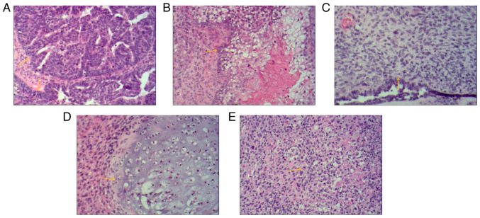
Figure 4.
Histological images of bilateral adnexal masses. Extensive metastases were observed throughout the pelvis and abdomen. Histological images of bilateral adnexal masses revealed the carcinosarcoma and sarcomatous elements of ovarian carcinosarcoma. (A) The main carcinomatous components included high-grade serous carcinoma. Tumor cells were arranged in glandular, papillary and solid patterns. The ducts were slit-like or irregular and poorly differentiated, and cells were arranged in sheets. The cells had obvious atypia, with large hyperchromatic pleomorphic nuclei, in which prominent nucleoli and numerous mitotic figures were present (yellow arrows). (B) Carcinomatous components included a small amount of squamous cell carcinoma. Tumor cells were arranged in nested, expansile, polygonal, paving stone-like patterns. Intercellular bridges were present and intracellular keratinization was observed in central cells. Nuclei were large, hyperchromatic or vacuolated with visible nucleoli and mitoses (yellow arrow). (C) Sarcomatous components contained fibrosarcoma. The tumor cells were spindle-shaped, with a cord-like distribution. Nuclei were hyperchromatic and mitotic bodies were present (yellow arrow). (D) Sarcomatous components contained a small amount of chondrosarcoma. Scattered chondrosarcoma was present in the cartilage lobules. The chondrocytes were located in the cartilage lacuna, with large hyperchromatic pleomorphic nuclei, in which binucleated cells and mitotic figures were present (yellow arrow). (E) Sarcomatous components also contained a small amount of rhabdomyosarcoma. The tumor cells were scattered in round and oval shapes. The cytoplasm was abundant and eosinophilic. Nuclei were eccentric and vacuolated with obvious nucleoli (yellow arrow) (H&E; magnification, x100).

