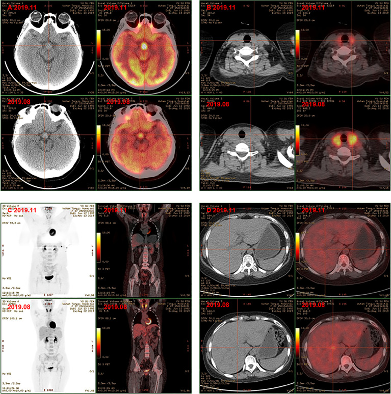
Figure 3.
PET-CT showed hyphothalamic nodule was thickened and the metabolism of hypothalamus was higher (A); the volume and metabolism of thyroid gland decreased (B); the range of primary lesions and the metabolism was significantly reduced, the metabolism of bilateral tonsils and bone marrow was similar to the previoud (C); metabolism of the multiple foci in the liver subsided (D) after chemotherapy.

