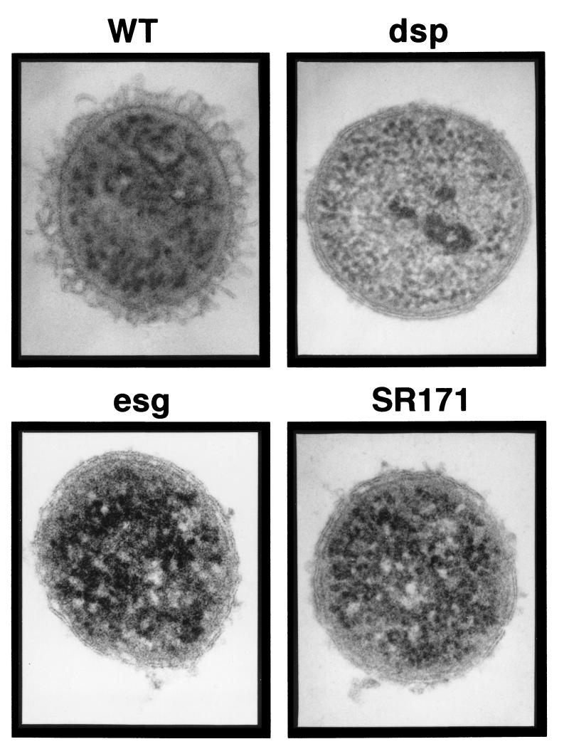
FIG. 7.
TEM of thin sections of freeze-substituted M. xanthus cells. Log-phase M. xanthus cells were removed from the growth medium and prepared for examination by TEM with the SFFS procedure as described in Materials and Methods. The M. xanthus strains examined were the wild type (WT) (DK1622) and the dsp (DK3468), esg (JD300), and SR171 mutants. The diameter of the cross sections of M. xanthus cells was 0.7 to 0.8 μm, and the thickness of the cell surface-associated layer observed with wild-type cells was approximately 80 nm.

