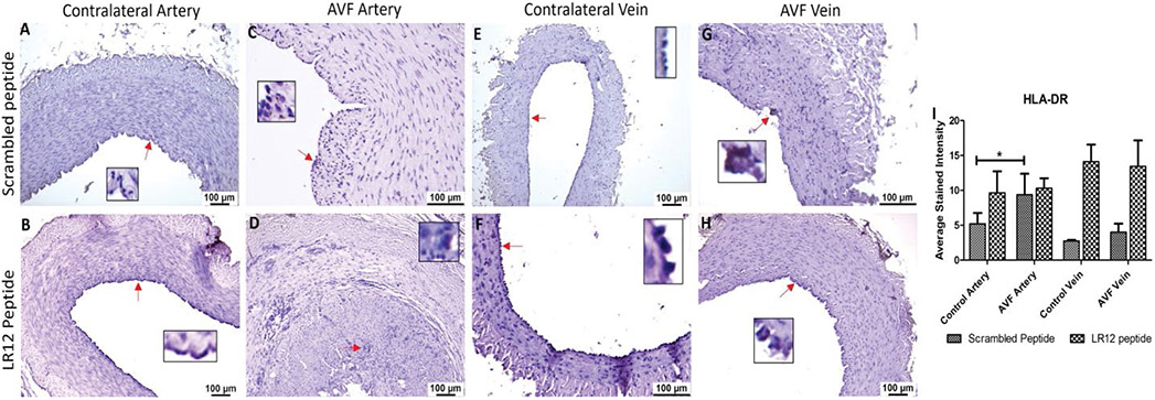
Figure 5:
Immunostaining for dendritic cells in femoral vessels involved in arteriovenous fistula (AVF) and contralateral femoral vessels in swine treated with triggering receptor expressed on myeloid cells (TREM)-1 inhibitor LR12 and scrambled peptide. Immunostaining for HLA-DR in contralateral femoral vessels (panels A, B, E, and F) and femoral vessels involved in AVF (panels C, D, G, and H), and average stained intensity (panel I). These images are represented images from all swine selected for this study. Inset shows higher magnification images of immune cells and red arrows show the positively stained immune cells. All data are presented as Mean ± SEM. A probability (p) value <0.05 was considered significant. *p<0.05.

