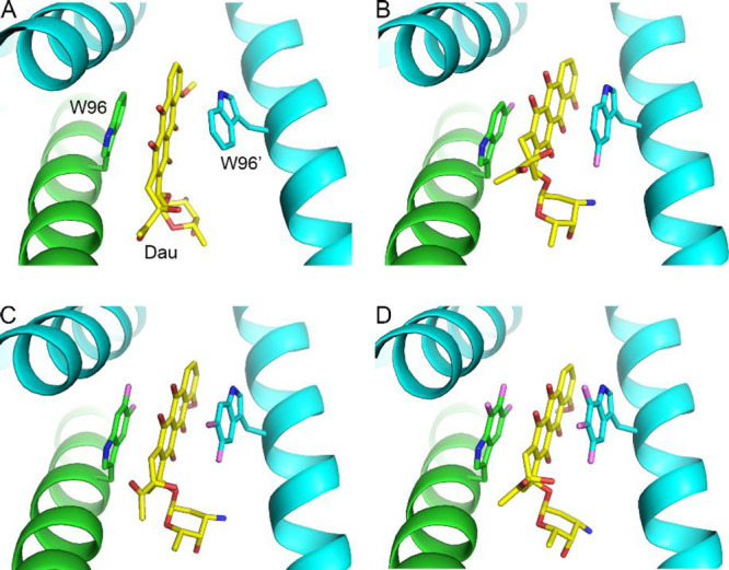
Figure 2.

Binding modes of daunomycin in the crystal structures of wild-type LmrR and the three LmrR W96 fluoro-substituted variants. (A) Wild-type LmrR-Dau complex (PDB entry 3F8F(19)), (B) 5FW-LmrR-Dau (PDB entry 7QZ6, this work), (C) 5,6diFW-LmrR-Dau (PDB entry 7QZ8, this work), (D) 5,6,7triFW-LmrR-Dau (PDB entry 7QZ7, this work). The two chains of the LmrR dimer are colored in cyan and green. Daunomycin is colored in yellow (carbons), red (oxygens), and blue (nitrogens).
