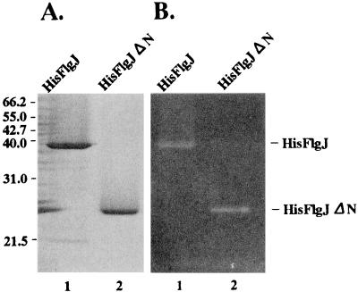
FIG. 3.
SDS-PAGE and zymogram analyses of the His-tagged FlgJ proteins. (A) The purified proteins were separated in an SDS–12% polyacrylamide gel. After electrophoresis, the gel was stained with 0.25% Coomassie brilliant blue. The molecular masses of the marker proteins are indicated in kilodaltons on the left. (B) The purified proteins were separated in an SDS–12% polyacrylamide gel containing M. lysodeikticus cells. After electrophoresis, the gel was treated as described in Materials and Methods for the zymogram analysis. Equal molar amounts of protein (54 pmol) were applied to each lane. The proteins used were His-FlgJ (lane 1) and His-FlgJΔN (lane 2).

