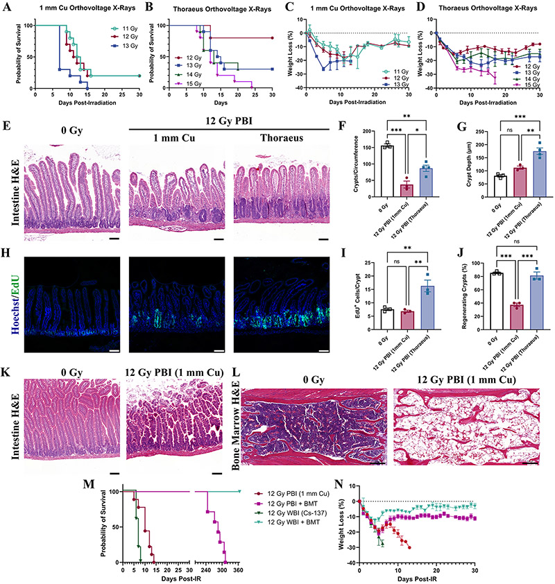
Figure 5: Response to Partial Body-Irradiation is Altered by Energy of Irradiation.
(A) Kaplan-Meier survival curves for mice receiving partial-body irradiation with either 1 mm Cu or (B) Thoraeus filtered orthovoltage X-rays. (C) Comparison of weight loss (% of starting body weight) following partial-body irradiation with either (C) 1 mm Cu or (D) Thoraeus filtration. (E) Representative Hematoxylin & Eosin (H&E)-stained intestinal sections from mice 4-days post-12 Gy partial-body irradiation. (Scale Bar 100 μm; n≥3/group.) (F) Total counts of crypts per circumference of intestinal cross-sections and (G) Crypt depth measurements 4-days post-irradiation analyzed by one-way ANOVA. (H) EdU incorporation was fluorescently detected (green) to assess cell proliferation in intestinal crypts 4-days post-12 Gy PBI (Scale Bar 100 μm; n≥3/group.) (I) EdU+ cells per intestinal crypt and (J) the percent of regenerating crypts (≥5 EdU+ cells/crypt) 4-days post-irradiation analyzed by one-way ANOVA. (K) Representative H&E-stained intestinal sections from moribund mice following 12 Gy partial-body irradiation. (Scale Bar 100 μm; n=5). (L) Representative H&E-stained bone marrow sections showing one sternebrae in moribund mice (n=5). (M) Kaplan-Meier survival curves from mice irradiated either with 12 Gy PBI (1 mm Cu) or 12 Gy WBI (Cs-137). Mice were either administered a PBS injection or bone marrow transplant 24 hours post-irradiation. (N) Weight loss as a percent of starting body weight for mice in the survival study. Data is represented as mean ± SEM. P<.05 (*), P<.01 (**), P<.001 (***).

