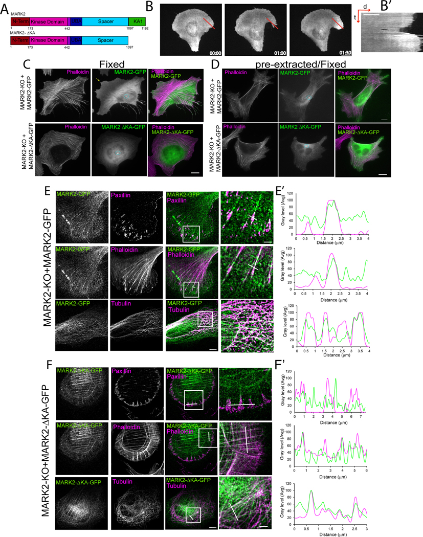
Figure 4: MARK2 association with the plasma membrane is required for leading edge advance and FA localization.
Imaging and analyses of U2OS cells (U2OS), or U2OS cells with MARK2 knocked out by CRSPR-cas9 (MARK2-KO) and MARK3 knocked down by siRNA (MARK-KO) or MARK2-KO cells expressing MARK2-GFP or MARK2-GFP lacking amino acids 1098-1192 (MARK2-ΔKAGFP). (A) Diagram of domains for MARK2 and MARK2-ΔKA lacking the KA1 membrane-targeting domain. (B) Time-lapse image series (time in min) and kymograph (B’) taken along the red line (right panel distance (d) and time (t)) of a cell expressing MARK2-GFP plated on a “crossbow”-shaped fibronectin island surrounded by non-adhesive substrate. Bar=10μm. (C-E) Localization of MARK2-GFP (C, D top, E) or MARK2-ΔKA-GFP (C, D, bottom, F) and staining of F-actin (C-F), paxillin (E, F) or tubulin (E, F) in fixed (C) or pre-extracted and fixed cells (D, E, F). White boxes: regions magnified at right (zoom), white lines: regions used for line scan analyses in (E’, F’). Circle and arrows in (C, D: MARK2 at the centrosome and a protrusion, respectively. Bars in (C, D, and E (left)= 5μm, (E, right)= 2μm. See also Figure S4, Video S5.

