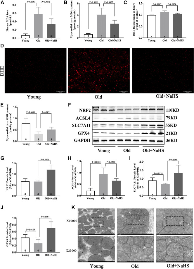
FIGURE 7.
H2S inhibited cardiomyocyte ferroptosis in aging rats. (A) MDA levels in the plasma of aging rats. (B) MDA levels in the myocardial tissue of aging rats. (C) Quantitative data on DHE-fluorescence in aging rats. (D) Representative DHE-fluorescence in the myocardial tissue sections of aging rats. (E) GSH levels in the myocardial tissue of aging rats. (F) Representative Western blots for NRF2, ACSL4, SLC7A11, and GPX4 expressions in the myocardial tissues of aging rats. GAPDH was used as the internal control. (G) The quantitative analysis for NRF2 expression in the myocardial tissue of aging rats. (H) Quantitative analysis for ACSL4 expression in the myocardial tissue of aging rats. (I) Quantitative analysis for the SLC7A11 expression in the myocardial tissue of aging rats. (J) Quantitative analysis for the GPX4 expression in the myocardial tissue of aging rats. (K) Representative images of transmission electron microscopy for mitochondria in the cardiomyocyte of aging rats. Results are means ± SEM. A p-value of <0.05 was considered significant.

