Figure 1.
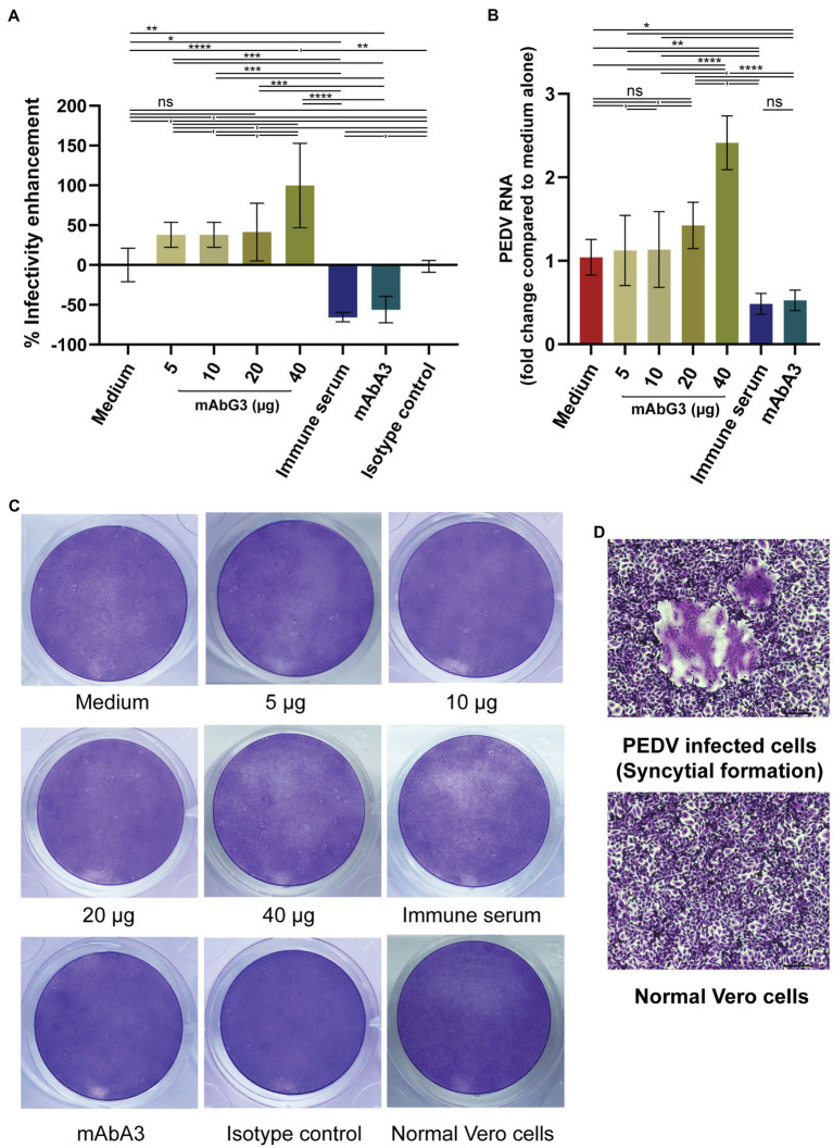
Enhancement of the PEDV infectivity mediated by the mAbG3. (A) The mAbG3 at the concentrations of 5, 10, 20, and 40 μg was mixed with 100 pfu of PEDV GII strain P70 and incubated for 1 h before adding to the confluent monolayers of Vero cells. Immune serum to PEDV S1 subunit (1:100) and mAbA3 (40 μg) served as positive neutralization controls, medium alone served as negative neutralization/enhancement control, and isotype-matched IgG1-kappa mAb served as irrelevant mAb control. After 1 h of incubation, the fluid in each well was removed; the cells were rinsed and covered with 2% CMC in DMEM supplemented with 2 μg/ml of TPCK-treated trypsin. Two days post-infection, cells were fixed, and the number of syncytial formations was counted and calculated to % enhancement of PEDV infectivity compared to the medium alone. Data of each bar graph are presented as the mean and standard deviation of the % mAbG3-mediated enhancement of PEDV infectivity from triplicate wells of each treatment. (B) Results of qRT- PCR for determination of PEDV RNA recovered from different treatment groups. Y-axis, fold changes of PEDV RNA in different treatment groups compared to medium alone; X-axis, various treatment groups. By one-way ANOVA: ns, not significantly different; *, p < 0.05; **, p < 0.01; ***, p < 0.001; ****, p < 0.0001. (C) Examples of the PEDV-mediated Vero cell CPE of different treatments. (D) Appearance of syncytial formation of PEDV-infected Vero cells (upper panel) compared with normal Vero cells (lower panel) at ×100 magnification of a light microscope.

