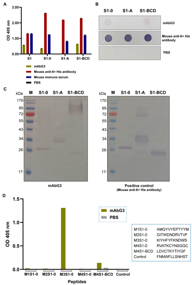
Figure 4.
Binding of mAbG3 to recombinant polypeptides S1-0, S1-A, and S1-BCD of the PEDV S1 subunit. (A) Indirect ELISA (OD 405 nm) of mAbG3 binding to the full-length recombinant S1 subunit and the S1 polypeptides S1-0, S1-A, and S1-BCD. Mouse anti-6× His antibody and mouse immune serum (1:10,000) served as positive binding controls, and PBS served as a negative binding control. (B) Binding of mAbG3 to S1-0, S1-A, and S1-BCD by dot ELISA. Mouse anti-6× His antibody (1:1,000) and PBS served as positive and negative binding controls, respectively. (C) Binding of mAbG3 to SDS-PAGE-separated S1-0, S1-A, and S1-BCD by Western blotting (left panel). Mouse anti-6× His antibody (1:1,000) was used as a positive control (right panel). (D) Binding of mAbG3 to M1S1–0, M2S1–0, M3S1–0, M4S1–0, M4S1-BCD, and control peptides by peptide binding ELISA. Y-axis, OD 405 nm; X-axis, peptides used as the test antigens; PBS served as control.

