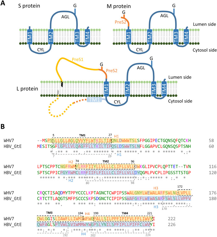
Fig. 1. Proposed surface protein topology and sequence alignment of WHV and HBV.
(A) Current purposed models for WHV and HBV S, M, and L protein topologies. The blue boxes indicate transmembrane helices (TM). The zigzag line in L protein indicates the N-terminal myristoylation. G, N-glycosylation; CYL, cytosolic loop; AGL, antigenic loop. (B) Sequence alignment of WHV7 and HBV genotype E (GtE) small surface proteins. The TMs were predicted by the TMHMM server, which are indicated by the dashed boxes with their designated position. The helix assignment from this study was presented in transparent colored boxes (orange, WHV; blue, HBV) with their designated position. Identical (*), conserved (:), and semiconserved (.) amino acids are indicated. Blue and purple indicate negatively or positively charged residues, respectively. Red indicates amino acids with hydrophobic side chains.

