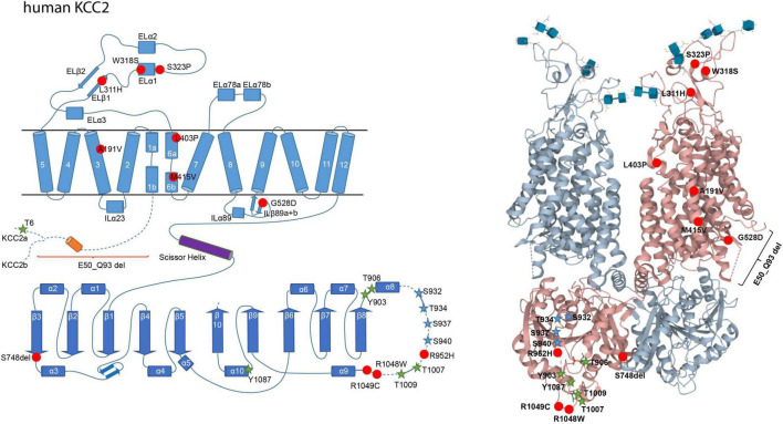
FIGURE 2.
Structural organization of human KCC2. 2-dimensional (left) and 3-dimensional (right) organization of human KCC2 according to Chi X. et al. (2020) (PDB: 6m23). KCC2 consists of 12 transmembrane domains (TMs) and two intracellular termini. A large extracellular loop is located between transmembrane domains 5 and 6 (EL3) and five N-glycosylation sites (blue cubes, left). Phosphorylation sites that increase KCC2 activity upon dephosphorylation are marked as green stars (Thr6 in KCC2a, Thr906, Tyr903, Thr1007, Thr1009, and Tyr1087). Phosphorylation sites that increase KCC2 activity upon phosphorylation are marked as blue stars (Ser932, Thr934, Ser937, Ser940). Human pathogenic variants of KCC2 associated with epilepsy, autism-spectrum disorder, and schizophrenia are depicted as red dots (Ala191Val, Leu311His, Trp318Ser, Ser323Pro, Leu403Pro, Met415Val, Gly528Asp, Arg952His, Arg1048Trp, Arg1049C, Ser748del). Annotation of amino acid residues is according to human KCC2b. The 3D reconstruction of KCC2 was generated using cryo-EM (Chi X. et al., 2020). 3D visualization was performed using Mol* Viewer in PDB (Sehnal et al., 2021).

