. 2022 Aug 5;11:e75863. doi: 10.7554/eLife.75863

Figure 2. Skeletal muscle-specific knockdown of Septin7 resulted in a severe phenotype.

(A) Images of tamoxifen-fed Cre- and Cre+ mice (both Septin7flox/flox) at the age of 4 months (see Figure 2—figure supplement 1). (B) Three-primer PCR for detecting the partial deletion of the Septin7 gene in different skeletal muscle types of Cre+ mice (Q: m. quadriceps femoris; B: m. biceps femoris; P: m. pectoralis) and the lack of deletion in samples prepared from BL6 control and Cre- mice. In samples originated from Cre- animals, the floxed exon 4, while in wild-type tamoxifen-fed mice the unmodified exon 4 is demonstrated. First and last lines in the ladder correspond to 200 and 300 bp, respectively. (C) Pooled data of the percentage of exon 4 deletion in different muscle types of Cre+ mice. 14 littermates (nine Cre+ and five Cre-) were examined from three litters. Here and in all subsequent figures, the rectangles in the box plots present the median and the 25 and 75 percentile values, while the error bars point to 1 and 99%. (D) Changes of body weight in Cre- (green triangle, n = 11) and floxed Cre+ (red square, n = 14) mice. Black solid line shows where the difference is statistically different (p<0.05 from t-test). Error bars show SEM. (E) Grip force normalized to body weight in Cre- (n = 4) and Cre+ mice (n = 7). Representative twitch (F) and tetanic force (G) transients in m. extensor digitorum longus (EDL). Peak twitch (H) and tetanic force (I) in EDL from Cre- (n = 7) and Cre+ (n = 11) mice. Calibration in panel (F): 1 mN and 50 ms; (G): 5 mN and 100 ms. **p<0.01, ***p<0.001 (from t-test). See also Figure 2—figure supplement 2. Main contractile proteins (actin and myosin) and L-type calcium channel distribution within single m. flexor digitorum brevis (FDB) myofibers isolated from Cre- and Cre+ animals were also investigated (see Figure 2—figure supplement 3).

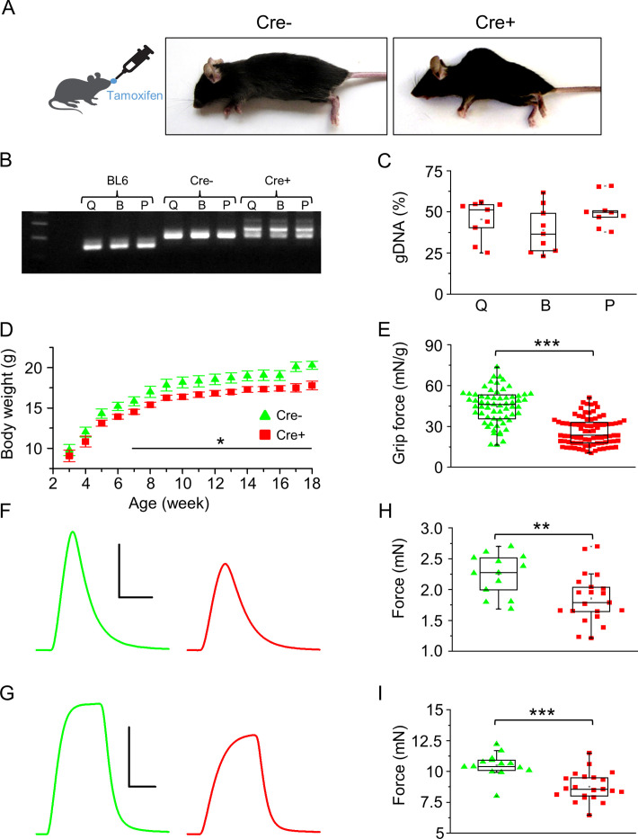
Figure 2—source data 1. Three-primer PCR was used to detect the partial deletion of the Septin7 gene in different skeletal muscle types of Cre+ mice (Q: m. quadriceps femoris; B: m. biceps femoris; P: m. pectoralis) and the lack of deletion in samples prepared from BL6 control and Cre- mice.
In samples originated from Cre- animals, the floxed exon 4, while in wild-type tamoxifen-fed mice the unmodified exon 4 is demonstrated. Relevant parts of the original gel images are presented in Figure 2B.

Figure 2.

Figure 2—figure supplement 1. Effects of Septin7 knockdown on the phenotype of Cre+ mice.

Figure 2—figure supplement 1.

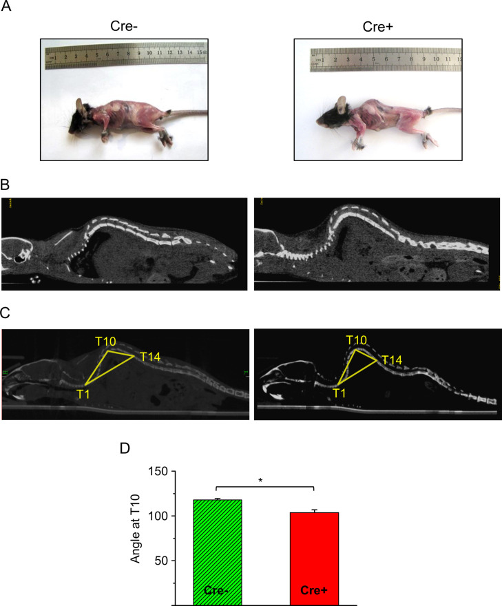
(A) Images of a tamoxifen-fed control (Cre-) and Cre+ mice. (B) Coronal CT image of a Cre-and Cre+ mouse. (C) Reconstruction of the whole backbone from CT images. A triangle was drawn to the thoracic 1st (T1), 10th (T10), and 14th (T14) vertebra, and the angle at T10 was determined. (D) Average of angles at T10 from three Cre- and three Cre+ mice (*p<0.05 from t-test). Error bars show SEM.
Figure 2—figure supplement 2. Effects of tamoxifen feeding on the phenotype of BL6 and Cre- mice.

Figure 2—figure supplement 2.

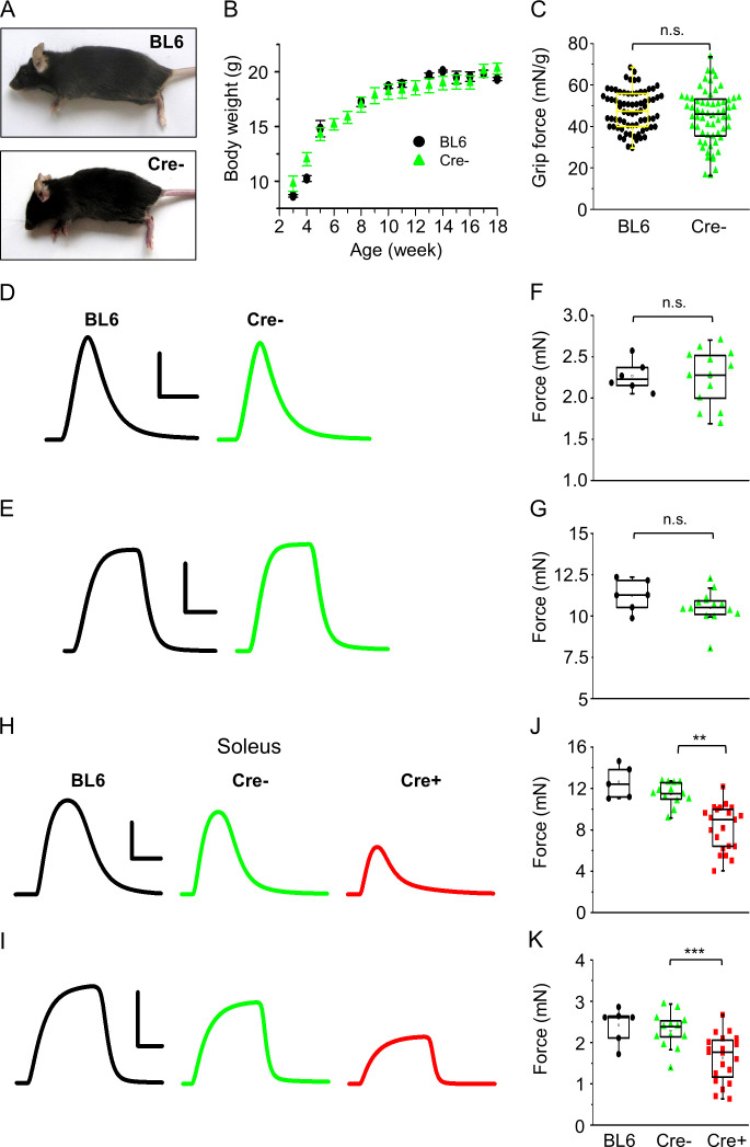
(A) Images of tamoxifen-fed BL6 and Cre- mice. (B) Body weight increase in BL6 (black circle, n = 3) and Cre- (green triangle, n = 11) mice. (C) Grip force normalized to body weight in BL6 (n = 3) and Cre- (n = 4) mice. Representative twitch (D) and tetanic force (E) transients in m. extensor digitorum longus (EDL). Peak twitch (F) and tetanic force (G) in EDL from BL6 (n = 3) and Cre- (n = 7) mice. Representative twitch (H) and tetanic force (I) transients in m. soleus (Sol). Peak twitch (J) and tetanic force (K) in Sol from BL6 (n = 3), Cre- (n = 7), and Cre+ (n = 11) mice. Calibration in panel (D) 1 mN and 50 ms; (E) 5 mN and 100 ms; (H) 1 mN and 100 ms; (I) 10 mN and 200 ms (**p<0.01, ***p<0.001 from t-test).
Figure 2—figure supplement 3. Expression and spatial distribution of actin, MYH4, and L-type calcium channels in single m. flexor digitorum brevis (FDB) muscle fibers isolated from Cre- and Cre+ animals.

Figure 2—figure supplement 3.

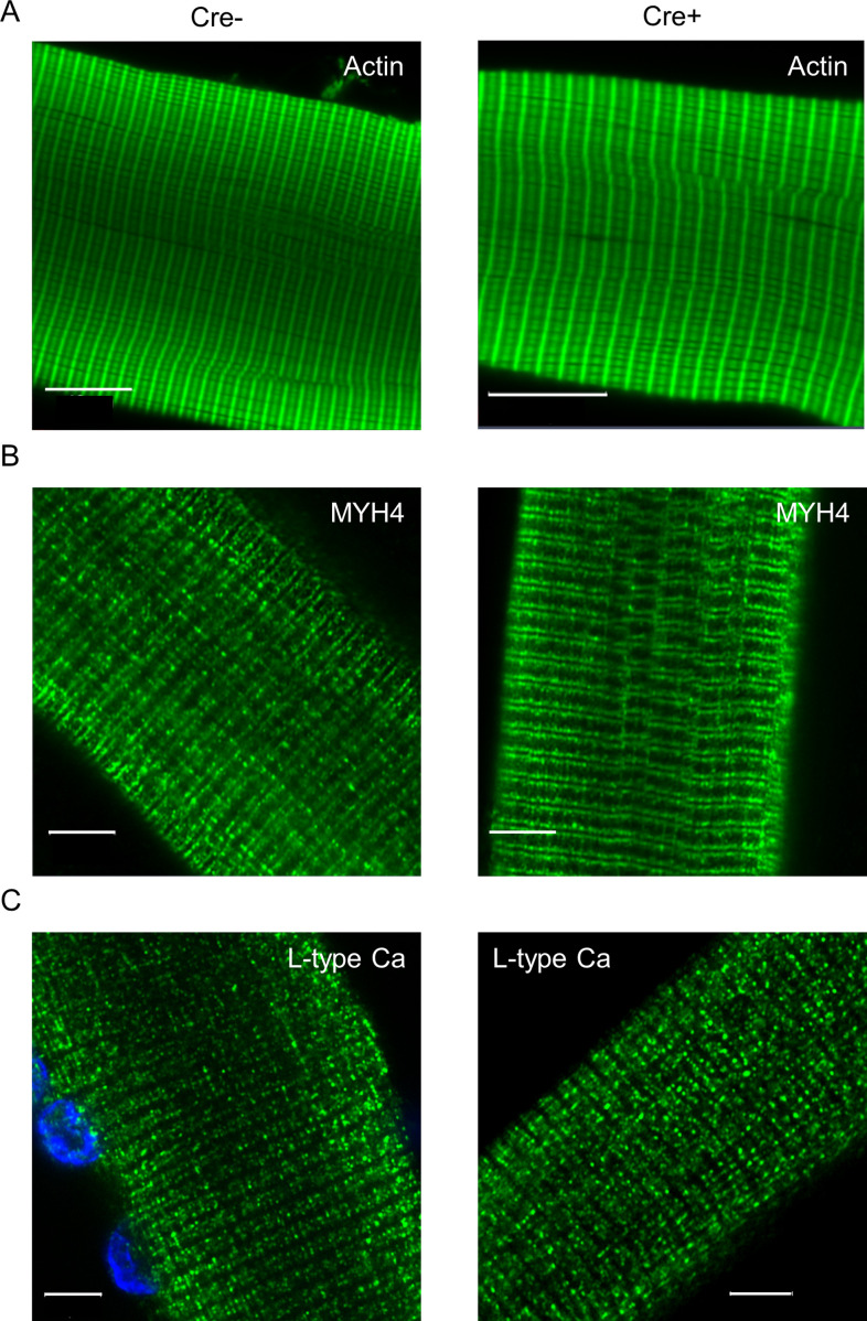
(A). Images represent actin-specific fluorescent labeling of single fibers isolated from Cre- and Cre+ animals. FITC-conjugated phalloidin was used for the detection of contractile protein actin. (B) MYH4-specific fluorescent labeling on isolated skeletal muscle fibers. (C) The presence and localization of L-type calcium channels were investigated on single muscle fibers isolated from Cre- and Cre+ mice. Cell nuclei (blue) are stained with DAPI. Scale bars represent 10 µm in panel (A) and 5 µm in panels (B) and (C).