. 2022 Jun 21;11:e77999. doi: 10.7554/eLife.77999

Figure 2. SERPINE3 gene loss pattern across 430 mammalian species.

(A) Left: Phylogeny of mammalian species investigated for the loss of SERPINE3 with mapped gene loss events indicated as red crosses. Branches of major clades are colored (Rodentia – brown, Chiroptera – orange, Eulipotyphla – blue, Afroinsectiphilia - red). The number of species investigated per clade is specified in parenthesis. For all loss lineages (red font), visual capability (classified as echolocating, fossorial/subterranean, vision as non-primary and primary sense) is displayed as pictograms at the right. Asterisk marks indicate species, where SERPINE3 evolved under relaxed selection but did not accumulate not more than one inactivating mutation. Right: The Serpin protein domain (Pfam) spans all eight protein-coding exons (boxes) of the intact human SERPINE3 gene (top). The secretion signal and the reactive core region are conserved in species with an intact SERPINE3 (Figure 2—figure supplements 9 and 10). Gene-inactivating mutations are illustrated for three clades, with stop codon mutations shown in black, frame-shifting insertions and deletions shown in red and mutated splice site dinucleotides shown between exons in red. Deleted exons are shown as red boxes. Insets show codon alignments with inactivating mutations in red font. RB - roundleaf bat, HB - horseshoe bat. (B) An ancestral deletion removed large parts of SERPINE3 in the tenrec lineage. UCSC genome browser (Lee et al., 2022) view of the human hg38 assembly showing the SERPINE3 locus and whole genome alignments between human and two tenrec species, visualized as a chain of co-linear local alignments. In these chains, blocks represent aligning sequence and double lines represent sequence between the aligning blocks that do not align between human and tenrec. A large deletion removed the first five protein-coding exons of SERPINE3 in both species. Shared breakpoints (gray box) indicate that the deletion likely represents an ancestral event in Tenrecidae.

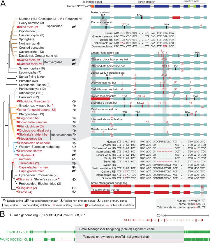
Figure 2—source data 1. SERPINE3 intactness in 447 mammalian assemblies.
Figure 2—source data 2. Verification of inactivating mutations in SERPINE3 with raw DNA sequencing reads.
Figure 2—source data 3. Expression of SERPINE3 remnants in public RNA-seq datasets.
Figure 2—source data 4. Analysis of relaxation or intensification of selection in SERPINE3 branches of interest.

Figure 2.

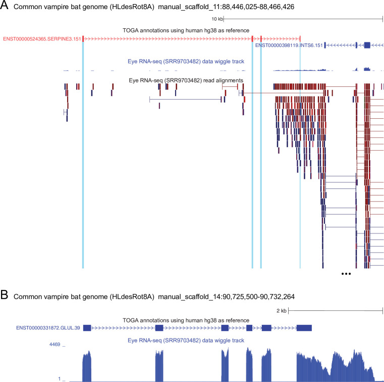
Figure 2—figure supplement 1. The remnants of SERPINE3 are not expressed in the common vampire bat.

Figure 2—figure supplement 1.

(A) UCSC genome browser screenshot of the common vampire bat (Desmodus rotundus) genome (Blumer et al., 2022) showing the four remaining exons of SERPINE3 (red: TOGA annotation, the other exons are deleted in this bat) and three coding exons of the neighboring INTS6 gene. Eye RNA-seq from D. rotundus (Sadier et al., 2018), shown as a wiggle track and read alignments, show that three SERPINE3 exons (blue highlight) are not expressed. The fourth SERPINE3 exon overlaps the 3' UTR of the neighboring INTS6 gene, which is located on the other strand. This explains why reads cover this SERPINE3 exon and its flanks, but there is not a single read that supports splicing of this exon to other SERPINE3 exons. In contrast, INTS6 exons are robustly expressed and show spliced alignments. (B) For comparison, the conserved GLUL gene, which is a key enzyme in glutamate detoxification, is highly expressed in eyes of the common vampire bat.
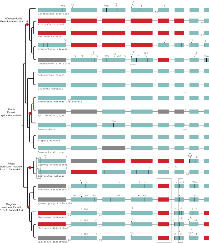
Figure 2—figure supplement 2. Gene-inactivating mutations support four independent losses of SERPINE3 in Afrotheria and Xenarthra (red dots).

Figure 2—figure supplement 2.

Those shared mutations are marked by boxes, which indicate the loss of SERPINE3 in the common ancestor of related species according to parsimony. For tenrecs (Microgale talazaci, Echinops telfairi) and elephant shrew (Elephantulus edwardii), the putative region of exon 4 was likely deleted after the shared frame-shifting mutations. SERPINE3 in sirenia (branches marked in red) evolve under relaxed selection. Coloring and legend as in Figure 2. Gray exons denote missing information.
Figure 2—figure supplement 3. Gene-inactivating mutations support a single loss of SERPINE3 in Pholidota.

Figure 2—figure supplement 3.

A partial deletion of coding exon 6 and three frame-shifting deletions in exon 7 are shared among all four species, indicating a single loss of SERPINE3 in the Pholidota lineage. Coloring and legend as in Figure 2.
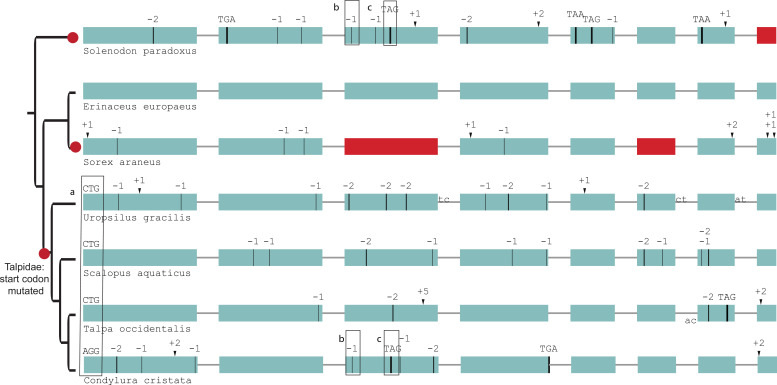
Figure 2—figure supplement 4. Gene-inactivating mutations support three independent losses of SERPINE3 in Eulipotyphla (red dots).

Figure 2—figure supplement 4.

The start codon is not intact in the four moles (a). Star-nosed mole (Condylura cristata) and Hispaniolan solenodon (Solenodon paradoxus) share two inactivating mutations in exon 3, although they are phylogenetically distant (b, c). Coloring and legend as in Figure 2.
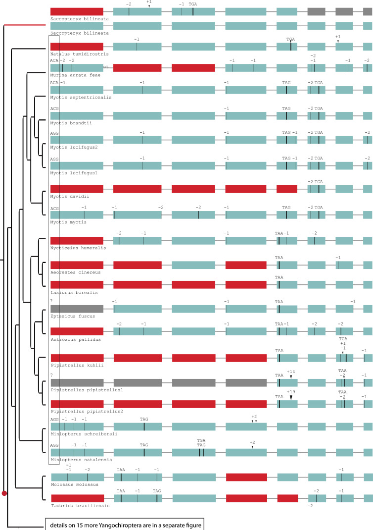
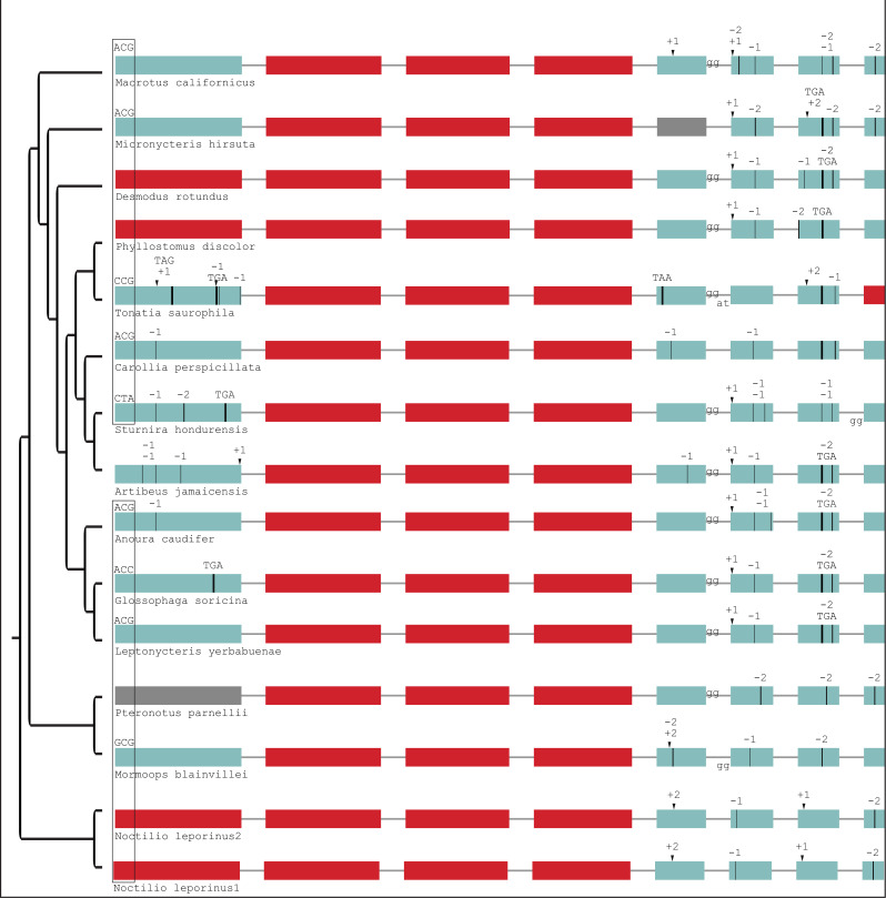
Figure 2—figure supplement 5. Gene-inactivating mutations support one loss of SERPINE3 in Yangochiroptera.

Figure 2—figure supplement 5.

The ancestral start codon is mutated in most Yangochiroptera (box). The most parsimonious explanation is a shared start codon mutation (red dot) in the ancestral lineage after split from the sac-winged bat (Saccopteryx bilineata). A back mutation to the regular start codon ATG likely occurred in Molossus molossus and Artibeus jamaicensis. Please see Figure 2—figure supplement 6 for gene-inactivating mutations of 15 more Yangochiroptera assemblies. The sac-winged bat has an intact copy of the gene that evolves under relaxed selection (red branch) in addition to a second copy of SERPINE3, which was independently inactivated. Coloring and legend as in Figure 2. Gray exons denote missing information.
Figure 2—figure supplement 6. Gene-inactivating mutations in additional Yangochiroptera.

Figure 2—figure supplement 6.

This is an extension of Figure 2—figure supplement 5. The start codon mutation shown is shared with most other Yangochiroptera (Figure 2—figure supplement 5). Coloring and legend as in Figure 2. Gray exons denote missing information.
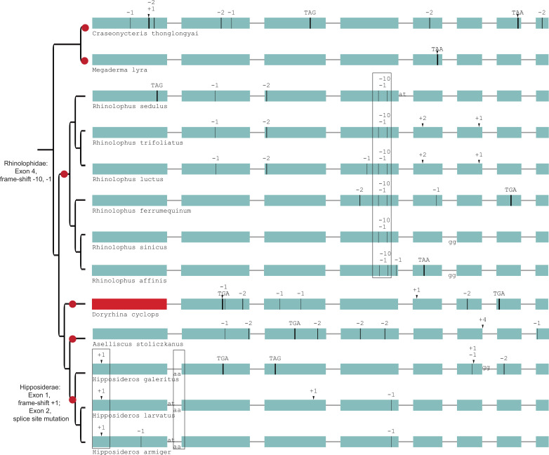
Figure 2—figure supplement 7. Gene-inactivating mutations support six independent losses of SERPINE3 in Yinpterochiroptera.

Figure 2—figure supplement 7.

Six independent inactivation events (red dots) occurred in SERINE3’s coding sequence in Yinpterochiroptera. All Rhinolophidae share frame-shifting mutations in exon 4 (boxed), while Hipposideridea share a frame-shifting insertion in exon 1 and a splice site mutation at exon 2 (boxed). Coloring and legend as in Figure 2.
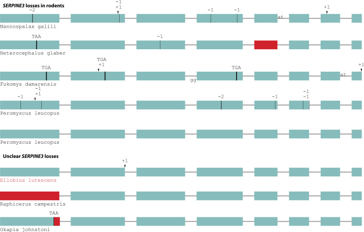
Figure 2—figure supplement 8. Independent gene-inactivating mutations in other mammals.

Figure 2—figure supplement 8.

A test for shifts in selective pressure revealed that SERPINE3 evolves under relaxed selection in the subterranean mole vole (Ellobius lutescens, marked in red), but not in the other cases of gene-inactivating mutations with unclear consequences (steenbok, okapi). A species-specific duplication with inactivation of one SERPINE3 copy occurred in white-footed mouse (Peromyscus leucopus). Coloring and legend as in Figure 2.
Figure 2—figure supplement 9. Conservation of the signal peptide in mammalian SERPINE3.

Figure 2—figure supplement 9.

The putative N-terminal signal peptide contains many hydrophobic residues in intact mammalian SERPINE3 and is predicted to guide secretion into extracellular space. We show the first 30 alignment columns. The sequence logo was generated with WebLogo (Crooks et al., 2004).
Figure 2—figure supplement 10. The hinge region and reactive core loop are conserved in intact mammalian SERPINE3.

Figure 2—figure supplement 10.

The numbers refer to human SERPINE3, where R369 likely is the scissile bond (P1)within the reactive core loop positions 366–372, (P4-P4’). The hinge region is located at positions 355–361. The logo was generated with WebLogo (Crooks et al., 2004).