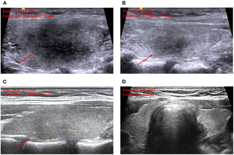
Figure 1.
Thyroid ultrasound imaging. Marked hypoechoic lesion on admission (A). Decreased hypoechoic lesion by half when the first follow-up on April 9 (B). Further decreased hypoechoic lesion when the second follow-up on May 21 (C). Disappeared hypoechoic lesion when the third follow-up on September 10 (D).

