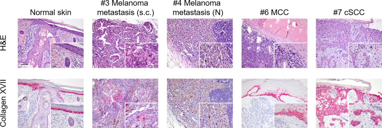
Figure 1.
Immunohistochemistry staining for collagen XVII (BP180) in malignant tumours of the skin. Using a rabbit monoclonal antibody (Abcam, clone: EPR14758) to collagen XVII (BP180), we stained formalin-fixed paraffin-embedded tissue sections of normal skin and tumour tissue available from a subgroup of our patient cohort. Upper line shows H&E stainings. Lower line shows staining for collagen XVII (BP180). Staining for collagen XVII in normal skin shows a physiological intercellular distribution in the basal layers of epidermal keratinocytes and following the adnexal structures into the deeper dermis. Nearly all tumour cells of the cutaneous squamous cells carcinoma (cSCC) showed strong positive staining for collagen XVII (patient #7). No positive staining was seen in melanoma cells of the nodal metastasis of patient #4 or in the Merkel cell carcinoma (MCC) cells of patient #6. In the latter, positive staining could only be detected in regions of physiological epidermis. Finally, weakly positive staining was found in melanoma cells of a subcutaneous metastasis of patient #3. Scale bar = 100 µm; insert scale bar = 25 µm.

