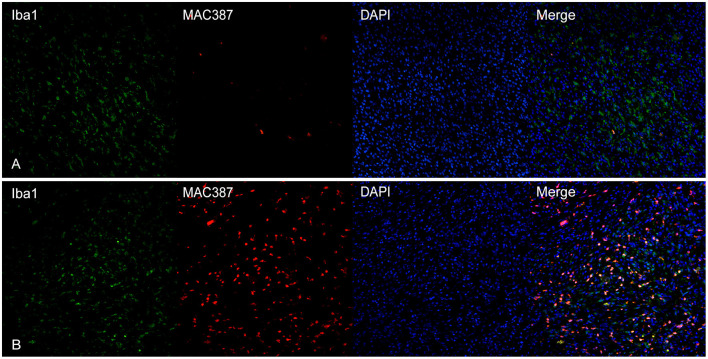
Figure 5.
(A) Canine, oral melanoma. Colocalization of Iba1 (green) and MAC387 (red). In melanomas, MAC387+cells were few and often did express also Iba1. (B) Canine, cutaneous melanoma. In areas near superficial ulceration, the number of cells co-expressing Iba1 and MAC387 was elevated. Nuclear counterstain was performed with DAPI.

