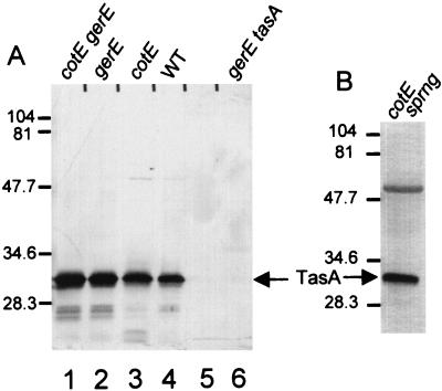
FIG. 3.
Western blot analysis (SDS–15% polyacrylamide gel) of TasA extracted from spores or a sporangial lysate. (A) Extracts of spores. Lanes: 1, cotEΔ::cat gerE36; 2, gerE36; 3, cotEΔ::cat; 4, wild type (WT); 5, empty lane; 6, gerE36 tasAΩpAGS08 (6). (B) Lysate of cotEΔ::cat sporangia (sprng) 5 h after initiation of sporulation. The arrows indicate the positions of TasA. Molecular masses are indicated in kilodaltons.

