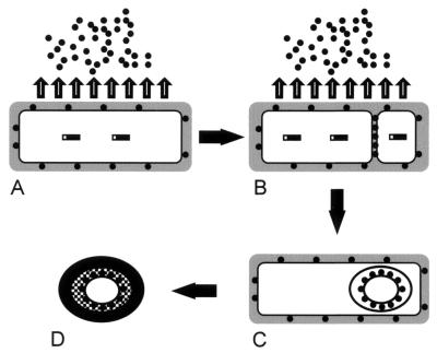
FIG. 9.
Model of the synthesis, processing, and translocation of TasA. (A) A cell that has committed to sporulation. (B) The same cell after the formation of the sporulation septum. (C) The same cell after engulfment of the forespore. (D) The mature spore, released after mother cell lysis. The hatched layer indicates the spore peptidoglycan, and the black layer indicates the coat. See Discussion for a detailed description. Solid rectangles, TasA; solid circles, secreted TasA; shaded area, cell wall.

