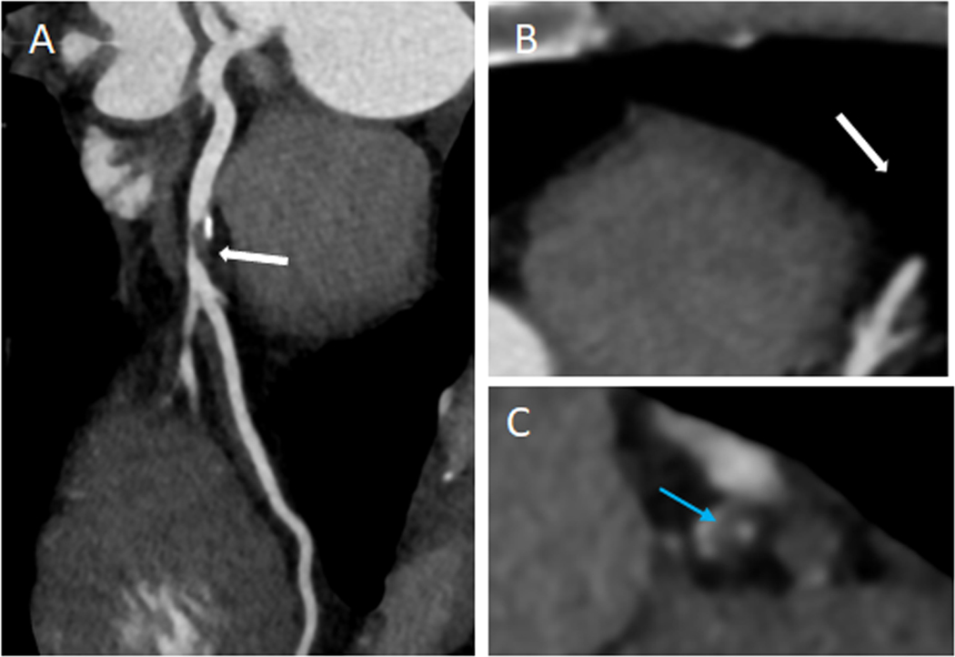
Figure 3.
High risk plaque features were a stronger predictor of MACE in women in PROMISE (aHR,2.41; 95% CI, 1.25–4.64). (A) Curved multiplanar reconstruction showing partially calcified plaque in the proximal LAD with evidence of positive remodeling (white-arrow). (B) Axial Image of the lesion. (C) Cross-sectional view of the plaque showing low attenuation plaque (blue arrow).

