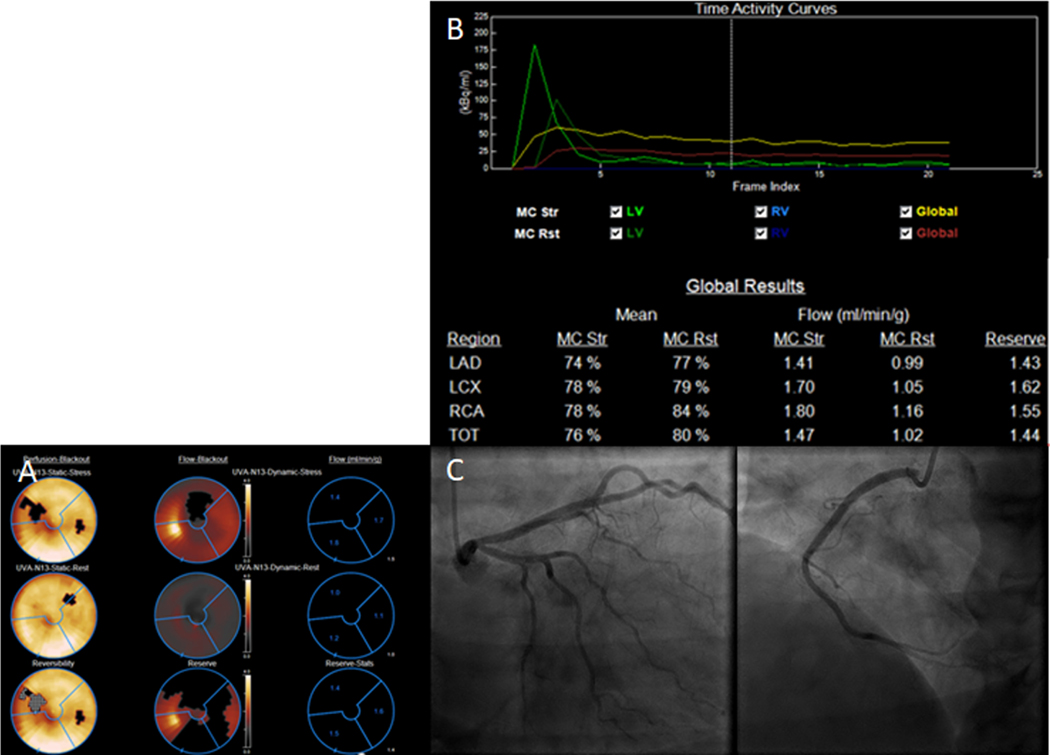
Figure 4.
Case of a 47 year-old female. (A) Stress PET study demonstrated a reversible perfusion defect of apical to mid anterior and inferoseptum segments. (B) Quantitative perfusion values show globally decreased perfusion during stress in the 3 coronary distributions, and globally decreased CFR in all 3 coronary distributions (C) Coronary angiogram demonstrated normal coronary arteries. The findings are consistent with INOCA/CMD

