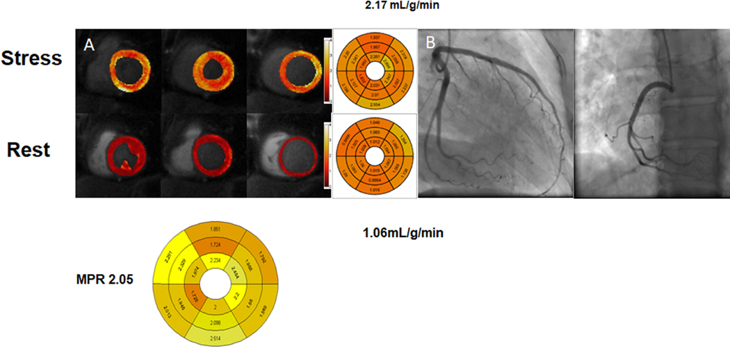
Figure 5.
Stress CMR quantification with MVD. In patients with risk factors for MVD, both MPR (2.21 [1.95,2.69] vs. 2.93 [2.763.19], p < 0.001) and stress myocardial perfusion (2.65 ± 0.62 ml/min/g, vs. 3.17 ± 0.49 ml/min/g p < 0.002) are reduced as compared to controls*.
A case of a 52 year old female with chest pain. (A) Adenosine-stress perfusion cardiac magnetic resonance imaging showing evidence of globally reduced coronary flow at stress, consistent with coronary microvascular dysfunction. Myocardial perfusion reserve index decreased at 2.05. (B) Coronary angiogram demonstrated normal coronary arteries.
Zorach B. Shaw PW, Bourque J, et al. Quantitative cardiovascular magnetic resonance perfusion imaging identifies reduced flow reserve in microvascular coronary artery disease. J Cardiovasc Magn Reson. 2018;20(1):14. Published 2018 Feb 22. doi: 10.1186/s12968-018-0435-1.

