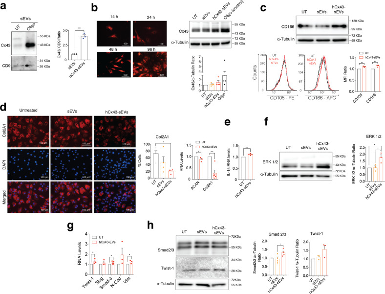
Fig. 2. Exosomal Cx43 induces dedifferentiation in target chondrocytes.
a Cx43 levels detected by WB in sEVs released by OA-derived chondrocytes treated with oligomycin (Oligo). n = 3, Student’s t test. b Cx43-positive sEVs increased Cx43 levels in target chondrocytes treated for 48 h. Oligomycin (Oligo) treatment (5 µM, 48 h) was used as positive control for Cx43 overexpression. n = 4. c WB and flow cytometry analysis shows increased levels of the mesenchymal markers CD105 and CD166 in OA-derived chondrocytes after 48-h treatment with sEVs derived from Oligo-treated chondrocytes. n = 3, Student’s t test. d Immunofluorescence for collagen type II (Col2A1) showed a significant decreased in OA-derived chondrocytes treated with hCx43-sEVs for 48 h (n = 4, one-way ANOVA). On the right, RNA expression of the main cartilage ECM markers ACAN and Col2A1 also showed a significant downregulation after a 48-h treatment with hCx43-sEVs (n = 3, Student’s t test). e IL-1β overexpression in OA-derived chondrocytes treated with sEVs derived from Oligo-treated chondrocytes. n = 3, Student’s t test. f MAPK/ERK pathway is activated in OA-derived chondrocytes after hCx43-sEVs treatment, as detected by higher ERK1/2 levels detected by WB. n = 3, one-way ANOVA. g Correlated to gene expression, protein levels of the EMT-transcription factors Smad2/3 (n = 4) and Twist-1 (n = 3) were increased in OA-derived chondrocytes after a 48-h treatment with hCx43-sEVs. Student’s t test. h The treatment with Cx43-positive sEVs was correlated with overexpression of several EMT-related transcription factors. n = 3, Student’s t test. Data are expressed as mean ± S.E.M., *P < 0.05, **P < 0.01, ***P < 0.0001.

