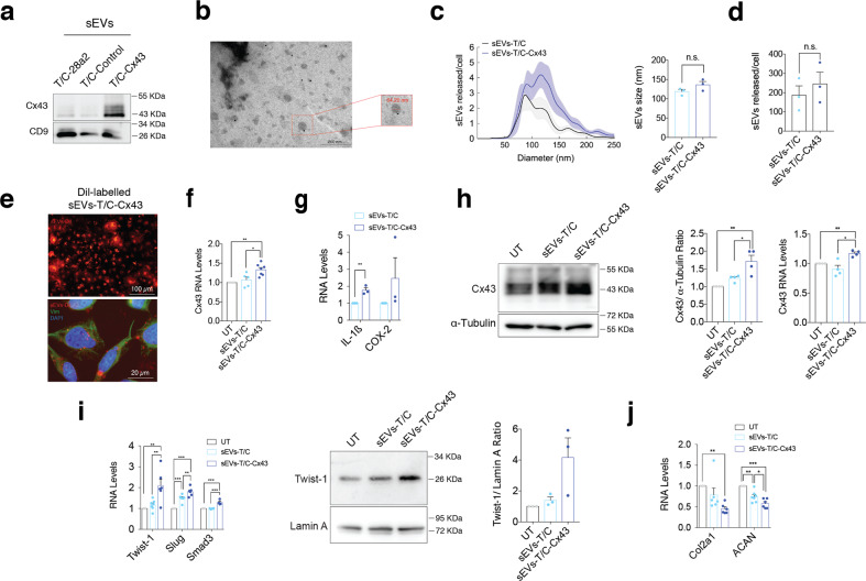
Fig. 3. sEVs derived from human osteoarthritic chondrocytes promote OA-phenotype in target chondrocytes.
a The human chondrocyte cell line T/C-28a2 overexpressing Cx43 released sEVs with higher Cx43 content as detected by WB. b Representative electron microscopy image of CD9-immunogold labelling from sEVs released by Cx43-overexpressing T/C-28a2 cells. c NTA analysis showing the average size of sEVs (in nm) derived from T/C-28a2 chondrocytes (sEVs-T/C) and Cx43-overexpressing T/C-28a2 chondrocytes (sEVS-T/C-Cx43), collected after 72 h. n = 3, Student’s t test. d NTA analysis of the number of sEVs released by Cx43-overexpressing T/C-28a2 cells and compared to non-transfected T/C-28a2 cells sEVs. n = 3, Student’s t test. e Red Dil-labelled sEVs isolated from Cx43-overexrpessing T/C-28a2 cells were detected in T/C-28a2 chondrocytes after 48-h treatment. Co-staining with vimentin antibody (green) and DAPI (blue) were performed to visualize the target cells. f Treatment of T/C-28a2 cells with high-Cx43 sEVs (sEVs-T/C-Cx43) for 48 h promoted Cx43 gene overexpression in target cells (n = 5–7, one-way ANOVA), as well as the expression of the inflammatory genes IL-1β and COX-2 (n = 3, Student’s t test) (g). h Treatment of T/C-28a2 cells with sEVs derived from Cx43-overexpressing T/C-28a2 chondrocytes (sEVs-T/C-Cx43) for 48 h increased both protein and gene expression levels in target chondrocytes. n = 4, one-way ANOVA. i When OA-derived chondrocytes were treated with sEVs-T/C-Cx43 for 48 h, EMT-markers gene (n = 5–7) and protein (n = 3) expression was upregulated, together with decreased gene expression levels of Col2A1 and ACAN (j, n = 6); one-way ANOVA. Data are expressed as mean ± S.E.M., *P < 0.05, **P < 0.01, ***P < 0.0001.

