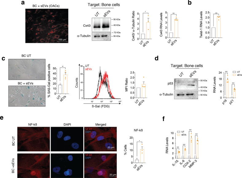
Fig. 7. sEVs secreted by OA-derived chondrocytes promote inflammation and senescence in bone cells.
a Red Dil-labelled sEVs isolated from OA-derived chondrocytes were detected in bone cells (BC) after 48-h treatment. Cx43 protein (middle; n = 3, Student’s t test) and RNA (right, n = 4; Student’s t test) levels were upregulated in bone cells treated with sEVs derived from OA-derived chondrocytes, as determined by western blot and RT-qPCR, respectively. b RNA levels of the EMT-transcription factor Twist-1 were also significantly upregulated in bone cells after a 48-h treatment with sEVs from OA chondrocytes. n = 4, Student’s t test. c Senescence-associated β-galactosidase activity (SA-β-Gal) was detected by microscopy (left; n = 4, Student’s t test) and flow cytometry (right; n = 4 Student’s t test) in bone cells treated for 48 h with sEVs derived from OA-derived chondrocytes. d Western blot analysis showed increase p53 protein levels in bone cells treated with sEVs isolated from OA-derived chondrocytes. On the right, gene expression levels of the senescence markers p16 and p21 were upregulated in BC after a 48-h treatment with sEVs from OA chondrocytes. n = 4, Student’s t test. e Treatment of BC with sEVs isolated from OA-derived chondrocytes for 48 h promoted NF-kβ nuclear translocation, as detected by immunofluorescence. n = 3, Student’s t test. f Overexpression of SASP factors IL-1β, IL-6, COX-2 and MMP-3 in BC treated for 48 h with sEVs isolated from OA-derived chondrocytes. n = 4, Student’s t test. Data are expressed as mean ± S.E.M., *P < 0.05, **P < 0.01, ***P < 0.0001.

