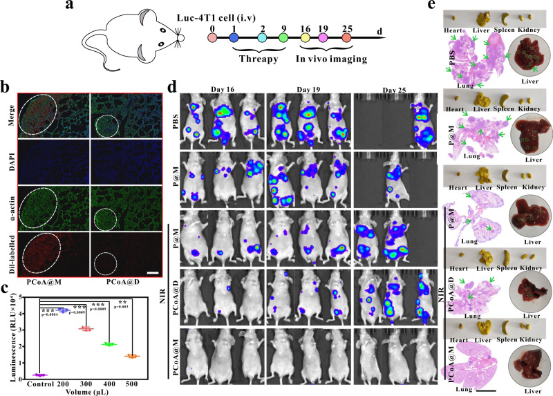
Fig. 9. Metastatic tumour therapy.
a Metastatic tumour model treatment process via injection of luc-4T1 cells into the tail vein. b α-Actin immunofluorescence imaging of lung tissues in mice with metastatic tumours after treatment with different Dil-labelled probes. Images are representative of three biologically independent mice. Scale bar: 200 μm. c Luminescence intensity of the luciferin reporter gene after luc-4T1 cells were incubated with different volumes of lysate. Data are presented as the means ± s.d. (n = 3 mice per group). Statistical differences were calculated using two-tailed Student’s t test, *: p < 0.05; **: p < 0.01; ***: p < 0.001. d Bioluminescence imaging at different times after the injection of different probes into mice bearing 4T1 lung metastases. e Bright field images of main organs fixed with Bouin’s trichrome (yellow) and original liver and HE staining of the corresponding lung tissues. Scale bar: 4 mm.

