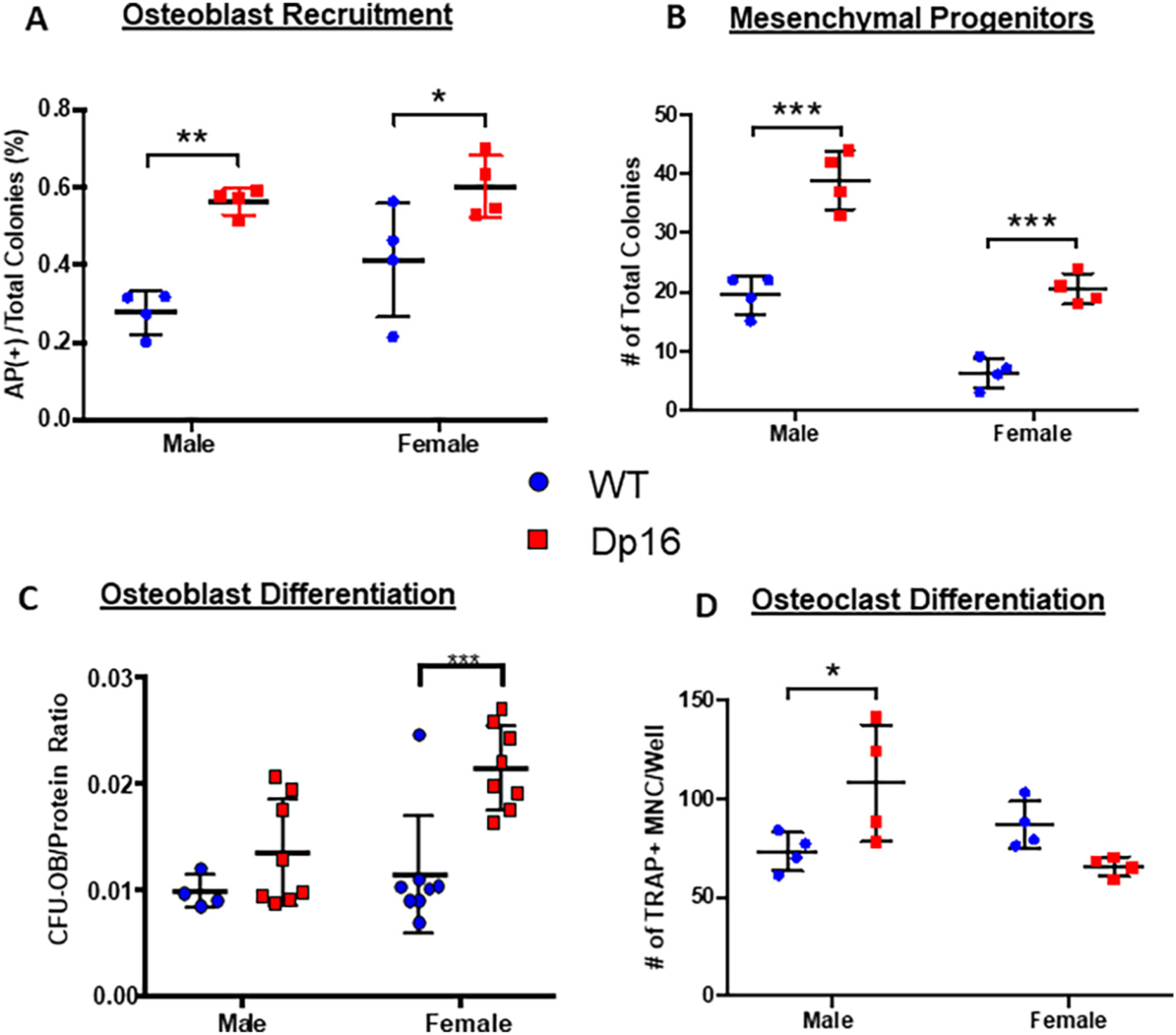
Fig. 3.

Dp16 DS ex vivo bone marrow cell differentiation. (A-C) Murine bone marrow cells from adult (3–4 months) WT (circles) and Dp16 DS (squares) male and female mice were cultured towards osteoblastogenesis. (A) Recruitment of cells into the osteoblast lineage was measured on d 10. The number of colonies staining positive for alkaline phosphatase (AP+) were counted and expressed as a percentage of the total number of colonies per well. (B) The total number of mesenchymal progenitors was determined in d 10 AP-stained and Fast green counter-stained dishes and both the number of AP+ colonies and the Fast green non-AP-stained colonies combined to express as total colonies per well. Number of mesenchymal progenitors was increased in both male and female Dp16 mice. (C) Mineralized osteoblastic colony-forming units (CFU-OB) were determined on d 28 by alizarin red staining, and the number of CFUOB normalized to the micrograms of protein content in each well. A significant increase in osteoblast differentiation capacity was observed in female Dp16 mice, but not male Dp16 mice. Data are representative of at least two similar experiments. p < 0.05 (*), p < 0.002 (**), or p < 0.001 (***) compared to WT gender-matched control. (D) Primary murine bone marrow cells were cultured for the development of osteoclasts. Cells were stained for TRAP activity, and the number of TRAP+ multinucleated cells (MNC) with 3 or more nuclei per well was counted. Data are representative of at least two similar experiments harvested on d 8–12. *p < 0.05.
