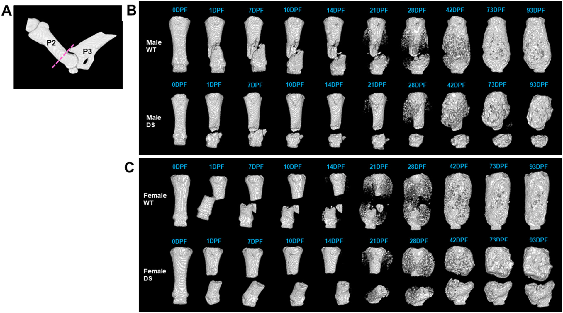
Fig. 4.

Time course of P2 Fracture Healing by in vivo microCT in WT and Dp16 DS mice. (A) MicroCT reconstruction of an intact P2 with the fracture plane (dashed line). In vivo microCT reconstructions of representative (B) male and (C) female WT and DS digits across the time course of fracture healing up to 93 days post-fracture (DPF). Normal fracture healing occurred in WT, with mineralization of the soft callus visible by 21 DPF, and fracture bridging by 42 DPF, when remodeling begins. Both male and female DS digits began mineralization by 21 DPF but did not successfully bridge by 42 DPF when WT digits were completely bridged and resulting in non-union fractures by 93 DPF.
