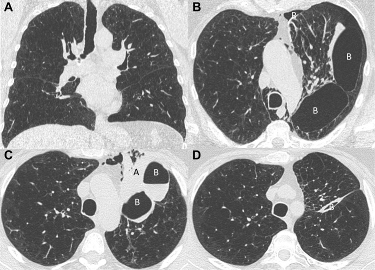
Figure 5.
Chest CT evolution (patient 5). (A) Baseline chest CT before endobronchial valves insertion in the left upper lobe (LUL). (B) Chest CT performed one day after valves insertion. We can see the hypoventilation of the LUL, presence of a pneumothorax (P) which did not require chest tube drainage, pneumomediastinum and the occurrence of two new bullae (B) between the upper and lower lobes. (C) Chest CT performed 5 days after valves insertion. There is a regression of the pneumomediastinum, increasing of the atelectasis (A) and the bullae (B) with an air–liquid level. (D) Chest CT performed 63 days after valves insertion with a partial atelectasis of the LUL and a nearly complete resolution of both bullae (B).

