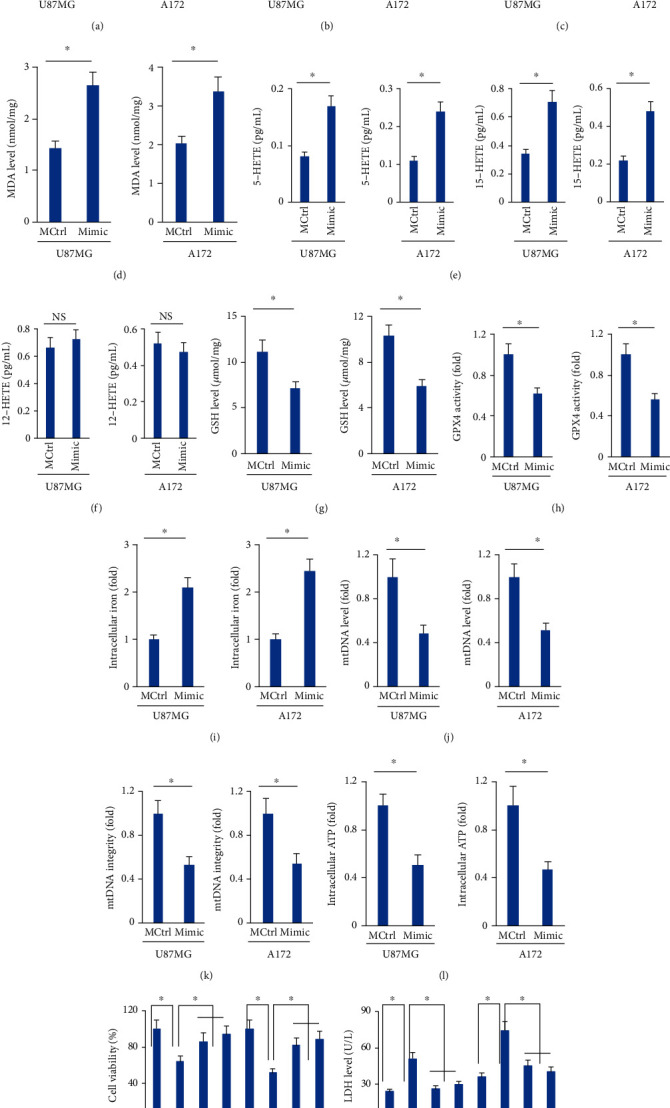
Figure 2.

The miR-147a mimic triggers ferroptosis of human glioblastoma cells. (a–b) miR-147a level in human glioblastoma cells treated with erastin or RSL3. (c) ROS level detected by DCFH-DA probe. (d) MDA level in cells treated with or without the miR-147a mimic. (e–f) 5-HETE, 12-HETE, and 15-HETE levels in the medium from cells treated with or without the miR-147a mimic. (g–h) GSH level and GPX4 activity in U87MG and A172 cells. (i) Relative level of intracellular iron. (j–k) Quantification of mtDNA content and integrity. (l) Quantification of intracellular ATP level. (m) Cell viability and LDH releases in the miR-147a mimic-treated human glioblastoma cells with Fer-1 or Lip-1 protection. N = 6 for each group. Data were expressed as the mean ± SD, and P < 0.05 was considered statistically significant.
