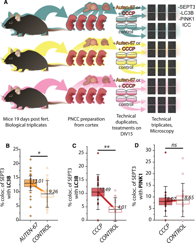
Fig. 5.
Experimental setup and results of colocalization analyses of septin-3 (SEPT3) due treatments. A Embryos were excised from three different dams to create biological triplicates of cultures. Embryonic cortices were isolated and homogenized to prepare cortical primary neuronal cell cultures. Two wells per culture were exposed to treatments (either AUTEN-67 or CCCP), two wells per culture were used as control. Cells were immunostained against SEPT3, LC3B and PINK1. Three images were captured and analyzed per well for every treatment/control condition. B–D Percentage comparison of colocalizing signals between treated and control cells. Means are marked with horizontal lines with values; boxes represent standard error of the mean (SEM). Flags indicate standard deviation (SD). In the figure legend, results are presented as mean ± SEM; SD. The p-values are of independent two-tailed Student’s t-test. B LC3B colocalizing SEPT3 in AUTEN-67 treatments, 13.01% ± 1.21%; 5.14%. Controls: 9.26% ± 1.1%; 4.68%. Significance: p = 0.0283. C LC3B colocalizing SEPT3, CCCP treatments: 10.49% ± 1.53%; 6.50%. Controls: 4.01% ± 1.06%; 4.38%. Significance: p = 0.0016. D PINK1 colocalizing SEPT3, CCCP treatment: 8.07% ± 1.52%; 6.46%. Controls: 8.65% ± 1.74%; 7.19%. Significance: p = 0.80

