. 2022 Apr 12;20(8):1284–1294. doi: 10.1158/1541-7786.MCR-21-0905

Figure 2.

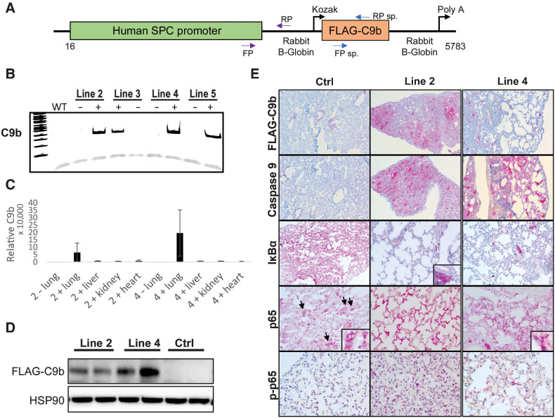
Figure 2. The SPC-driven C9b expression in mouse lung epithelium activates NF-κB pathway. A, A schematic diagram of a C9b transgene construct driven by human SPC promoter. Among the C9b transgene positive founder lines (B, PCR), line 2 and 4 C9b+ mice showed high levels of C9b transgene expression in lungs both for mRNA (C, real time rt-PCR, relative ratio of C9b/GAPDH) and protein (D, Western blot). E, Representative images of IHC staining with indicated antibodies (10x). Note a Flag-tagged C9b expression in lung epithelium, a decreased level of IκBα and an increased level of p-p65 in line 2 and line 4 mice compared with control mice. Insets (40x) in p65 images show the exclusion of p65 from nuclei in control but not in C9b mice. Inset in IκBa shows its strong positivity in immune aggregates.

The SPC-driven C9b expression in mouse lung epithelium activates NF-κB pathway. A, A schematic diagram of a C9b transgene construct driven by human SPC promoter. Among the C9b transgene positive founder lines (B, PCR), line 2 and 4 C9b+ mice showed high levels of C9b transgene expression in lungs both for mRNA (C, real time rt-PCR, relative ratio of C9b/GAPDH) and protein (D, Western blot). E, Representative images of IHC staining with indicated antibodies (10x). Note a Flag-tagged C9b expression in lung epithelium, a decreased level of IκBα and an increased level of p-p65 in line 2 and line 4 mice compared with control mice. Insets (40x) in p65 images show the exclusion of p65 from nuclei in control but not in C9b mice. Inset in IκBa shows its strong positivity in immune aggregates.