Figure 5.
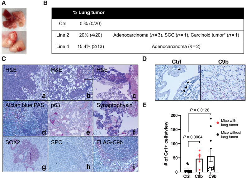
C9b mice develop lung tumors with aging. Macroscopic (A) and microscopic lung tumors observed in C9b mice over 21 m old of age are summarized in B. *, 1 mouse developed a carcinoid tumor and adenocarcinoma. C, Histologic analyses including H&E (a–c), Alcian Blue-PAS (d), and IHC staining with indicated antibodies (e–i) of lung tumors identified adenocarcinoma (a, d, g–i), squamous cell carcinoma (SCC; b and e), and carcinoid tumors (c and f). Images are taken at 10x magnification. Inset in b is at 40x magnification. D and E, Gr1+ cells (D, IHC) in the lung per low power field (20x, 3 views per mouse) were counted in control (n = 9) and C9b mice with (red, n = 5) or without (n = 11) lung tumors (E). Data are means ± SEM. Adjusted P values are determined by ANOVA with Tukey multiple comparison test.

