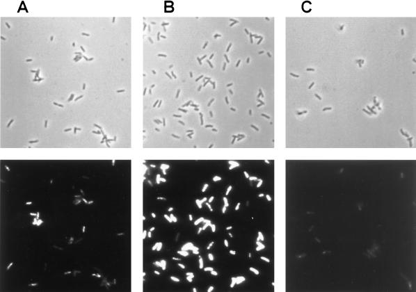
FIG. 3.
Detection of groEL mRNA in S. typhimurium LT2 cells. Cells were treated as described in Materials and Methods. Phase-contrast photomicrographs (top panels) and epifluorescence photomicrographs (bottom panels) of the same fields are shown. (A) Mid-log phase cells. (B) Cells heat shocked at 45°C for 15 min. (C) Cells heat shocked at 45°C for 15 min and RNase-treated prior to the in situ RT-PCR.

