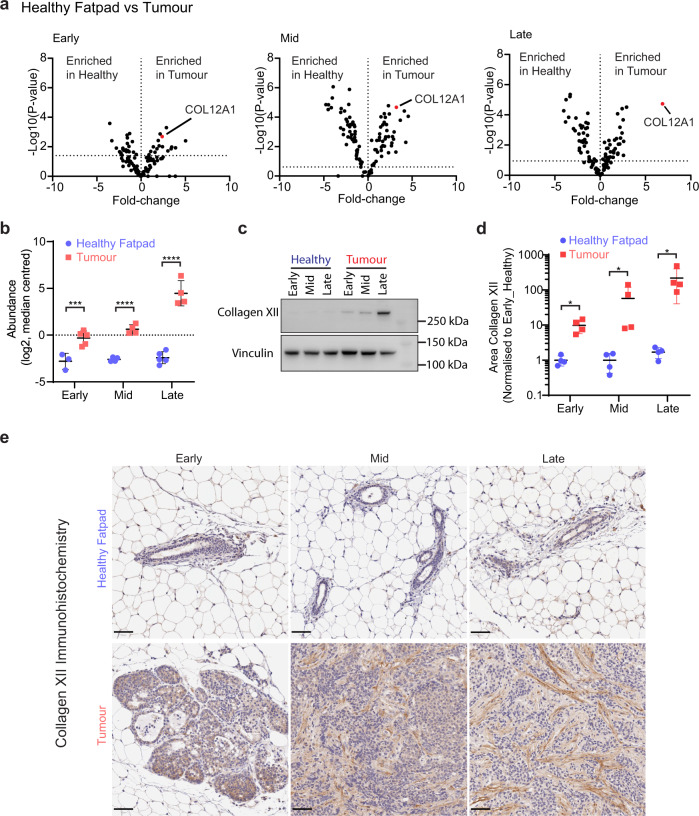
Fig. 2. Collagen XII is upregulated in breast tumours as the disease progresses.
a Volcano plot of differentially abundant matrisome proteins from LC-MS/MS in matched healthy fatpad or mammary tumour at the early, mid or late stages highlighting collagen XII elevation in mammary tumours at each stage. Multi-sample t test with FDR correction. Data derived from n = 4 mice for the tumour group at mid stage and n = 5 mice for all other groups. b Collagen XII protein abundance in tumour and matched healthy fatpad tissue, quantified by LC-MS/MS (log2 transformed, median centred, collagen XII was detected in n = 25 of 30 samples corresponding to tissue samples from n = 3 mice in the healthy early stage group, n = 4 mice in the healthy mid, tumour mid and tumour late stage groups and n = 5 mice from the healthy late and tumour early stage groups). ***p = 0.0003, ****p < 0.0001 One-way ANOVA with Holm–Sidak multiple comparison test. Data are presented as mean ± SD. c Temporal profile of collagen XII protein expression in healthy fatpad and primary tumours via western blot. Representative of n = 3 biologically independent samples. d Quantification of collagen XII immunohistochemistry (IHC) staining of staged tumours and matched healthy fatpad tissue. Average of 5× fields of view per tumour in n = 4 mice per group. *p = 0.029 Mann–Whitney U-test (two-sided). Data are presented as mean ± SD. e Representative histological images of collagen XII staining quantified in d). Scale bar = 50 µm from n = 4 biologically independent samples. Source data are provided in the Source data file.

