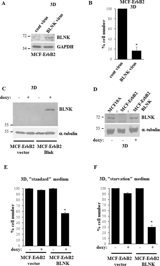
Fig. 4. Downregulation of BLNK is required for ErbB2-induced 3D growth of breast cancer cells.

A MCF-ErbB2 cells infected with the control (cont virus) or the BLNK-encoding MMLV (BLNK virus) were cultured detached from the ECM for 24 h (3D) and assayed for BLNK levels by western blotting. GAPDH was used as a loading control. B MCF-ErbB2 cells treated as in A were detached from the ECM for 72 h (3D) and counted. The number of cells detected in the control sample was designated as 100%. C Indicated cell lines were cultured detached from the ECM (3D) for 48 h in the absence (−) or in the presence (+) of 5 ng/ml doxycycline (doxy) and assayed for BLNK expression by western blotting. α-tubulin was used as a loading control. D Indicated cell lines were cultured detached from the ECM (3D) for 6 h in the absence (−) or in the presence (+) of 5 ng/ml doxycycline (doxy) and assayed as in C. A short vertical black line was used to indicate that lanes were removed from the image and separate parts of an image were joined together. E Indicated cell lines were cultured for 72 h as in C in the ″standard″ medium (Hybri-Care medium supplemented with 10% Fetal Bovine Serum) used for culturing these cells in all experiments except for F and counted. The number of cells detected in the control sample was designated as 100%. F Cells cultured in the ″starvation″ medium which represented the Hybri-Care medium medium supplemented with 0.1% Fetal Bovine Serum and diluted 2.5-fold with HBSS medium were assayed as in E. The data in B, E, F are the average of the three independent experiments plus the SD. *p-value < 0.05.
