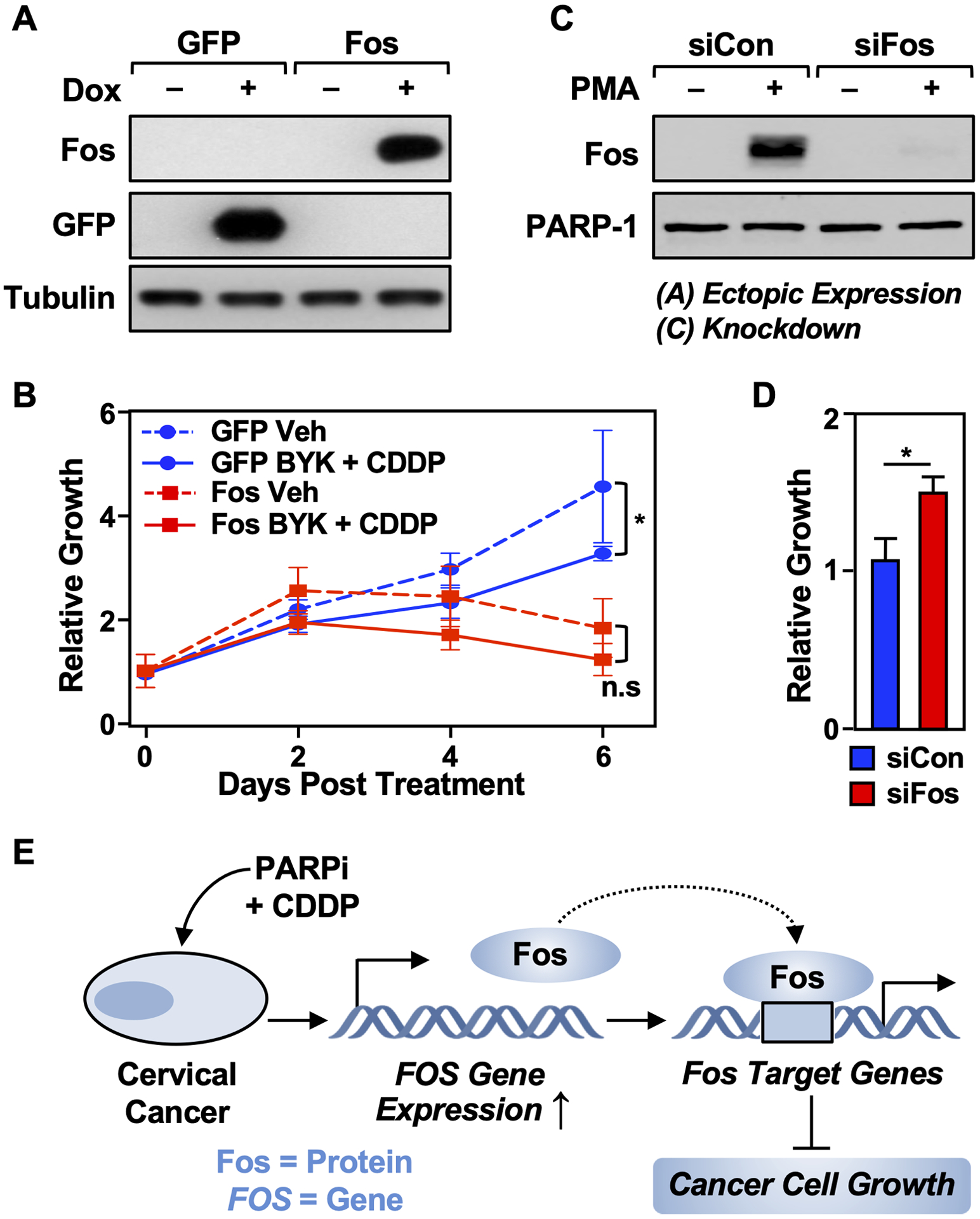
Figure 6. Fos expression drives the cytotoxic effects of BYK + CDDP.

(A) HeLa cells were engineered to ectopically express either Fos or GFP. Immunoblots show the doxycycline (Dox)-inducible expression of Fos and GFP as indicated. β-Tubulin was used as a loading control. The cells were treated with 1 mg/mL of Dox for 16 hours.
(B) Ectopic expression of Fos in HeLa cells leads to reduced growth of the cells. The cells were treated with Dox as in (A) and vehicle (DMSO) or BYK + CDDP as indicated. Cell growth was quantified after 0, 2, 4, or 6 days using a crystal violet assay. Each point represents the mean ± SEM (n = 3; Sidak’s multiple comparison test; * p < 0.05; n.s. is not significant at p < 0.05).
(C) Immunoblots showing the siRNA-mediated knockdown of Fos in HeLa cells. The cells were treated with PMA to induce Fos expression for visualization.
(D) Depletion of Fos in HeLa cells leads to increased growth of the cells. HeLa cells were treated with Fos siRNA as shown in (C) and cell growth was quantified after 2 days using a crystal violet assay. Each point represents the mean ± SEM (n = 3; Student’s t-test; *< 0.05).
(E) Model showing the inhibition of cervical cancer growth by co-treatment with PARP inhibitor and cisplatin via the induction of FOS expression. See the text for details.
