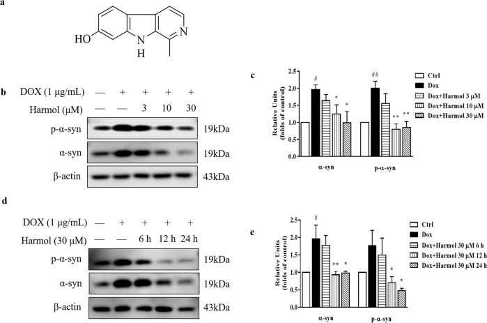
Fig. 1. Harmol degrades the α-syn.
a Chemical structure of harmol. b Tet-on inducible PC12 cells were treated with 1 µg/mL DOX for 24 h, then 3, 10, and 30 µM harmol were added for 24 h. The expression of p-α-syn and α-syn were determined by western blot. Representative blots are shown. c Relative intensity was normalized to that of β-actin. Data are presented as the mean ± SEM from three independent experiments. #P < 0.05 and ##P < 0.01 vs. the control (0.1% DMSO). *P < 0.05 and **P < 0.01 vs. the DOX. d After induction with DOX for 24 h, PC12 inducible cells were incubated with harmol (30 µM) for 6 h, 12 h, and 24 h. The expression of p-α-syn and α-syn were determined by western blot. Representative blots are shown. e Relative intensity was normalized to that of β-actin. Data are presented as the mean ± SEM from three independent experiments. #P < 0.05 vs. the control (0.1% DMSO). *P < 0.05 and **P < 0.01 vs. the DOX.

