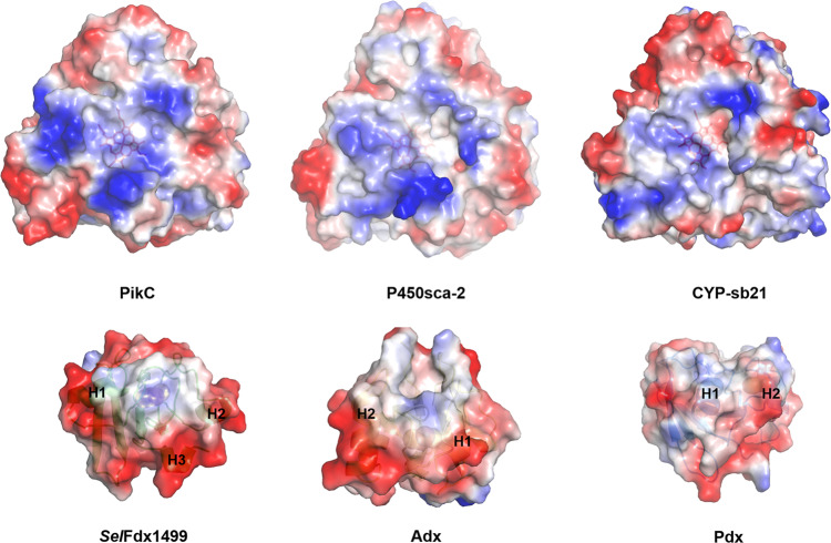
Fig. 4. Electrostatic surface analysis of P450s and Fdxs.
Positively and negatively charged surfaces are colored in blue and red, respectively. The electrostatic surfaces of Fdxs with Helix1 (H1), Helix2 (H2), or Helix3 (H3) are marked. The six proteins are PikC/YC-17 (PDB ID: 2VZ7), P450sca-2 (predicted by Alphafold2), CYP-sb21 (substrate-free, PDB ID: 6M4S), SelFdx1499 (predicted by Alphafold2), Adx (PDB ID: 1CJZ), and Pdx (PDB ID: 1PDX).

