Figure 2.

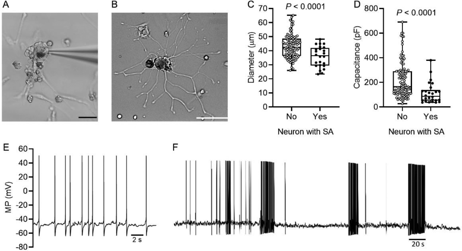
Spontaneous activity (SA) occurs preferentially in smaller dissociated human DRG neurons. (A) Human DRG neuron during whole cell patch recording one day after dissociation, showing the patch pipette, neurites, probable satellite glial cells (and possibly other cell types), and cellular debris. Calibration bar is 50 μm. (B) Example of a recorded neuron with extensive growth of neurites by the second day after dissociation. Calibration bar is 50 μm. (C) Neurons with SA had smaller soma diameter (unpaired t test) as well as lower membrane capacitance (D) (Mann-Whitney U test) compared with other neurons sampled. (E) SA recorded from a dissociated human DRG neuron illustrating the low-frequency, irregular discharge pattern and irregular, subthreshold DSFs between APs. (F) The only sampled neuron to show a bursting pattern during SA. The pattern of bursting and burst durations were irregular throughout the recording (6 min shown).
