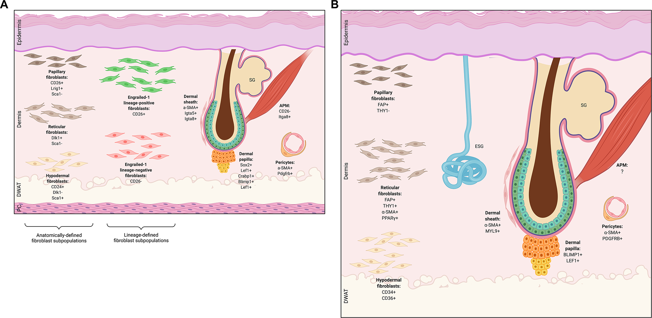
Figure 1: Differences in the architecture of mouse versus human skin.

A) Depiction of mouse skin structure, which includes a panniculus carnosus (PC) muscle layer under the hypodermis. B) Depiction of human skin structure. Human skin differs from mouse skin in that it is thicker overall, has sparser hair follicles (HF), and contains eccrine sweat glands (ESG) which are not found in mice outside the skin of the paws. For both mouse and human skin schematics, various mesenchymal subpopulations and characteristic markers are shown, including microanatomically defined fibroblast lineages (papillary, reticular, hypodermal fibroblasts), functionally defined fibroblast lineages (Engrailed-1 lineage-positive and lineage-negative fibroblasts), HF-associated fibroblasts (dermal sheath, papilla, arrector pili), and pericytes. SG, sebaceous gland; DWAT, dermal white adipose tissue; APM, arrector pili muscle.
