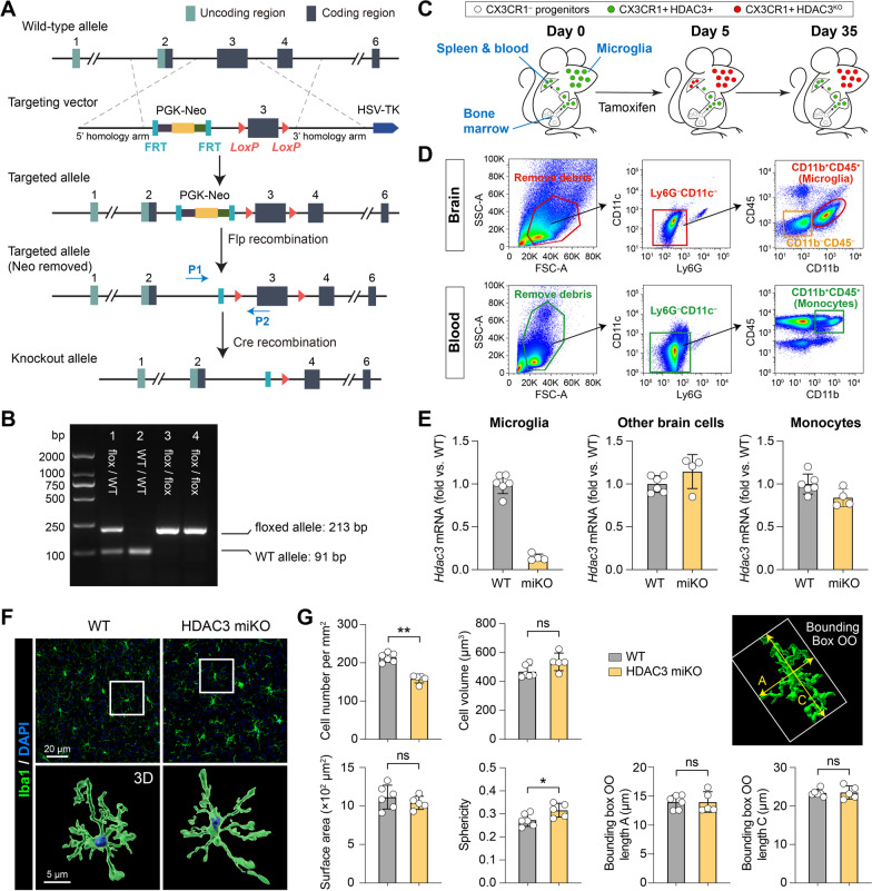
Fig. 1.
Generation and characterization of microglia-specific HDAC3 knockout mice. A Targeting strategy to generate HDAC3LoxP mice with LoxP-flanked exon 3 of the Hdac3 gene. In the presence of Cre, the LoxP sites recombine and delete the floxed region of Hdac3. P1 and P2, genotyping primers. B PCR genotyping demonstrating generation of heterozygous mice (lane 1) and subsequent breeding to obtain homozygous mice containing the LoxP sites on both alleles (lanes 3 and 4). Using primers flanking the 5′ LoxP site, the floxed allele runs at a weight of 213 bp, whereas the wild-type (WT) allele has a weight of 91 bp. C Pulse knockout strategy to restrict tamoxifen-induced HDAC3 KO to microglia. Peripheral CX3CR1+ cells are continuously replaced from CX3CR1– progenitor cells in the bone marrow. HDAC3 is deleted 5 days after tamoxifen treatment in both microglia and peripheral CX3CR1+ cells. After another 30 days, peripheral CX3CR1+ cells are replenished from bone marrow progenitors by new CX3CR1+ cells that have not undergone recombination. Therefore, only microglia are HDAC3 KO, and most peripheral monocytes express HDAC3. D FACS gating strategy for brain microglia (CD11b+CD45+), other brain cells (CD11b–CD45–), and blood monocytes (CD11b+CD45+). E Quantitative PCR was performed 30 days after tamoxifen treatment on FACS-sorted cells to verify the deletion of HDAC3 in microglia but not in other brain cells or blood monocytes. n = 6 (WT) and 4 (HDAC3 miKO) mice. F, G The number and morphology of microglia were assessed in the brain of HDAC3 miKO mice and WT control mice after Iba1 immunostaining and 3D rendering of images. n = 6 mice (237 cells) for WT. n = 5 mice (140 cells) for HDAC3 miKO. Shown are the mean ± SD. *p < 0.05, **p < 0.01. ns no significant difference

