Fig. 2.
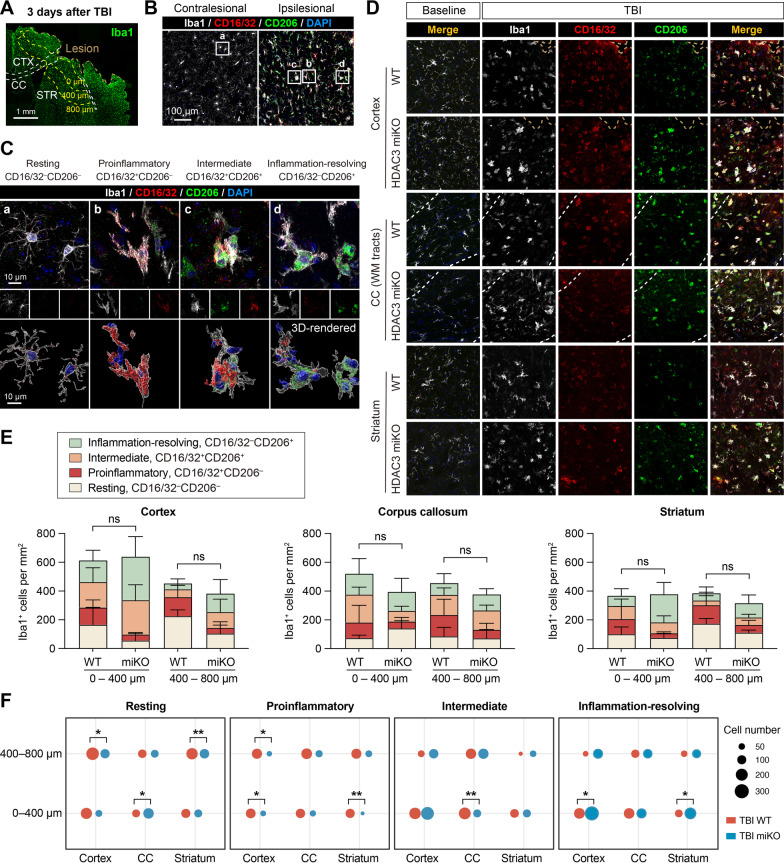
HDAC3 miKO enhances inflammation-resolving responses of microglia after TBI. HDAC3 miKO mice and WT control mice were subjected to TBI induced by controlled cortical impact. The phenotype of microglia was examined 3 days after TBI by triple-label immunostaining of CD16/32, CD206, and Iba1. A Iba1 immunofluorescence in the ipsilesional brain hemisphere illustrates the boundary of the TBI lesion and the peri-lesion areas in the cortex (CTX), corpus callosum (CC), and striatum (STR) where images in B–D were taken from. B Triple-label immunosignal of Iba1, CD16/32, and CD206 in the peri-lesion striatum and in the corresponding region in the non-injured contralesional cortex. Rectangles, areas enlarged in C. C Images taken under high magnification demonstrate 4 typical phenotypes of microglia based on their expression of CD16/32 and CD206: resting (a), proinflammatory (b), intermediate (c), and inflammation-resolving (d). Lower panels: images 3D-rendered by Imaris. D Representative images taken from the peri-lesion cortex, CC white matter (WM) tracts, and striatum 3 days after TBI or taken from the corresponding regions in baseline control brains. See Additional file 1: Fig. S2 for images of baseline controls under individual color channel. E The number of Iba1+ cells under each of the four phenotypic categories was counted in the peri-lesion cortex, CC and striatum, 0–400 μm and 400–800 μm from the lesion boundary. ns no significant difference in total Iba1+ cells between HDAC3 miKO and WT mice. Shown are the mean ± SD. F The numbers of Iba1+ cells in the 4 phenotype categories were illustrated in dot plots, where the area of a dot represents the number of cells. n = 6 (WT) and 5 (HDAC3 miKO) mice. *p < 0.05, **p < 0.01 HDAC3 miKO vs. WT

