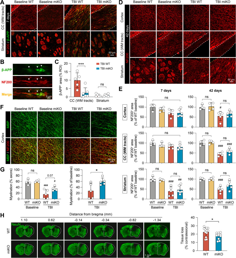
Fig. 5.
HDAC3 miKO improves short-term and long-term integrity of white matter after TBI. Immunofluorescence staining was performed at 7 and 42 days after TBI or non-injury control procedures (baseline controls) to assess brain injury and white matter integrity in HDAC3 miKO and WT mice. A–C Axonal injury was assessed 7 days after TBI using NF200 and β-APP double-label immunostaining. A Representative images taken from the white matter-enriched corpus callosum (CC) and striatum in the ipsilesional brain hemisphere. Dashed line, the boundary of CC. B β-APP immunosignal in the shape of classic axonal bulbs and varicosities (arrows) was observed after TBI, suggesting axonal damage. C Summarized data on β-APP-immunopositive areas. D Axonal integrity was assessed 42 days after TBI by NF200 immunostaining. E Summarized data on NF200-immunopositive areas in the ipsilesional cortex, CC and striatum 7 and 42 days after TBI, expressed as percentages of the WT baseline group. F Myelin integrity in the peri-lesion cortex was assessed 42 days after TBI using MBP and NF200 double-immunostaining. G Summarized data on the degree of myelination (areas immunopositive for both MBP and NF200), expressed as percentages of myelinated axons to total axons (left panel), or as percentages to baseline controls (right panel). n = 4 (baseline) or 5–6 (TBI) per group. H The volume of chronic brain tissue loss was measured 42 days after TBI on coronal brain sections immunostained for the neuronal marker NeuN. Dashed line, the relative area of the contralesional hemisphere to illustrate ipsilesional tissue loss. n = 12 mice per group. Shown are the mean ± SD. #p < 0.05, ##p < 0.01, ###p < 0.001 TBI vs. baseline. *p < 0.05, ***p < 0.001 HDAC3 miKO vs. WT. ns no significant difference

