Fig. 5.
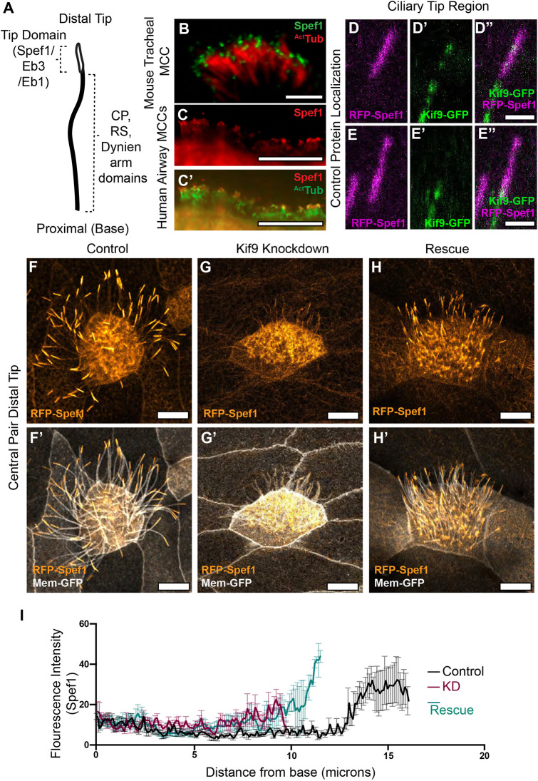
Kif9 contributes to distal tip integrity. (A) Schematic of proximal to distal patterning of the motile cilium. Tip domain schematized with proteins known to localize to that domain, as well as proteins localizing to the rest of the axonemes. (B) Images of immunostaining of Spef1 in mouse tracheal multiciliated cells (MCCs). Spef1 in green, acetylated tubulin (ActTub) in red. (C,C′) Images of immunostaining of human airway multiciliated cells. Spef1 in red, acetylated tubulin in green. (D–E″) Confocal images of RFP–Spef1 (magenta) and Kif9–GFP (green) colocalization in the distal tips of the cilium. (F–H′) Confocal images of RFP–Spef1 (orange) and Membrane–GFP (gray) in control (F,F′), Kif9 MO-injected (G,G′), and rescue (H,H′) embryos. (I) Plot showing RFP–Spef1 fluorescence intensity as a function of axoneme length. Note the higher intensity for RFP–Spef1 at the distal end of control but not Kif9-KD axonemes. Note too that both axoneme length and distal Spef1 enrichment are partially rescued by re-expression of Kif9 (N=45 control axonemes; 45 Kif9-KD axonemes; 42 rescue axonemes; data compiled from 15 embryos across three independent experiments). Scale bars: 1 µm in B; 10 µm in C,C′,F–H′; and 2 µm in D–E″.

