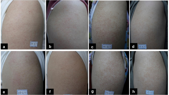
Fig. 2.
The comparison of clinical photographs at the baseline and at the last follow-up visit. a, b, e, f A mild-moderate KP patient’s photographs at the baseline (a, e) and at the last follow-up visit (b, f) for the laser side (a, b) and the control side (e, f). c, d, g, h A severe KP patient’s photographs at the baseline (c, g) and at the last follow-up visit (d, h) for the laser side (c, d) and for the control side (g, h)

