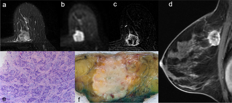
Fig. 4.
A 45-year-old woman with triple-negative tumour of the left breast. a Axial fat-suppressed T2-weighted image shows an 18-mm, heterogeneously hyperintense round mass with non-circumscribed margins in the upper inner quadrant of the left breast. b Axial diffusion-weighted imaging (b-value = 1,000 s/mm.2) shows peripheral high signal intensity and central hypointensity. c Axial and (d) sagittal post-contrast T1-weighted subtracted images show a corresponding irregular round mass with rim enhancement. e On cut surface, the lesion is round shaped with smooth margins. f Histologic examination confirms the diagnosis of infiltrating carcinoma of “no special type”

